Papillary muscle
The papillary muscles are muscles located in the ventricles of the heart. They attach to the cusps of the atrioventricular valves (also known as the mitral and tricuspid valves) via the chordae tendineae and contract to prevent inversion or prolapse of these valves on systole (or ventricular contraction).[1] The papillary muscles constitute about 10% of the total heart mass.
| Papillary muscle | |
|---|---|
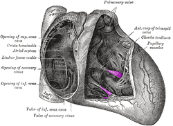 Interior of right side of heart. Papillary muscles labeled in purple. | |
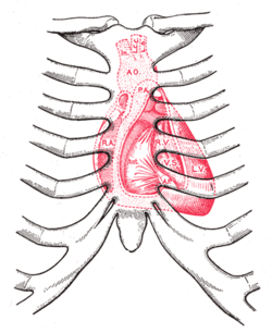 Diagram showing relations of opened heart to front of thoracic wall. Ant. Anterior segment of tricuspid valve. A O. Aorta. A.P. Anterior papillary muscle. In. Brachiocephalic artery (Innominate). L.C.C. Left common carotid artery. L.S. Left subclavian artery. L.V. Left ventricle. P.A. Pulmonary artery. R.A. Right atrium. R.V. Right ventricle. V.S. Ventricular septum. | |
| Details | |
| Identifiers | |
| Latin | musculus papillaris |
| MeSH | D010210 |
| TA | A12.1.00.022 |
| FMA | 12154 |
| Anatomical terms of muscle | |
Structure
There are five total papillary muscles in the heart; three in the right ventricle and two in the left. The anterior, posterior, and septal papillary muscles of the right ventricle each attach via chordae tendineae to the tricuspid valve. The anterolateral and posteromedial papillary muscles of the left ventricle attach via chordae tendineae to the mitral valve.[2]
Blood supply
The mitral valve papillary muscles in the left ventricle are called the anterolateral and posteromedial muscles.[3]
- Anterolateral muscle blood supply: left anterior descending artery - diagonal branch (LAD) and left circumflex artery - obtuse marginal branch (LCX)
- Posteromedial muscle blood supply: right coronary artery - posterior interventricular artery (RCA)
The posteromedial muscle ruptures more frequently because it only has one source of blood supply, hence RCA occlusion can cause papillary muscle rupture.[3]
Function
The papillary muscles of both the right and left ventricles begin to contract shortly before ventricular systole and maintain tension throughout.[1] This prevents regurgitation—backward flow of ventricular blood into the atrial cavities—by bracing the atrioventricular valves against prolapse—being forced back into the atria by the high pressure in the ventricles.[1]
Clinical significance
Papillary muscle rupture can be caused by a myocardial infarct, and dysfunction can be caused by ischemia. Both complications may lead to worsening of mitral regurgitation.[4]
Additional images
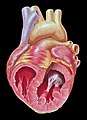 Opened chambers of the heart displaying papillary muscles and chordae tendinae
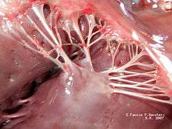 Papillary muscles and chordae tendinae
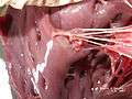 Papillary muscles and chordae tendinae
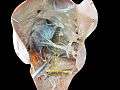 Papillary muscles. Deep dissection.
See also
References
- Moore KL, Dalley AF, Agur AM (2007). Clinically Oriented Anatomy (3rd ed.). Baltimore: Lippincott Williams & Wilkins. pp. 92, 94. ISBN 978-0-7817-6274-8.
- Netter's Atlas of Human Anatomy, plates 216B and 217A
- Fradley, M. G.; Picard, M. H. (7 March 2011). "Rupture of the Posteromedial Papillary Muscle Leading to Partial Flail of the Anterior Mitral Leaflet". Circulation. 123 (9): 1044–1045. doi:10.1161/CIRCULATIONAHA.110.984724. PMID 21382906.
- Elizabeth D Agabegi; Agabegi, Steven S. (2008). Step-Up to Medicine (Step-Up Series). Hagerstwon, MD: Lippincott Williams & Wilkins. p. 40. ISBN 0-7817-7153-6.
External links
- Anatomy photo:20:19-0106 at the SUNY Downstate Medical Center} — "Heart: The Right Atrioventricular (Tricupsid) Valve" (anterior, posterior, septal papillary muscles)
- Anatomy photo:20:26-0105 at the SUNY Downstate Medical Center — "Heart: The Left Atrioventricular (Mitral) Valve" (anterior, posterior papillary muscles)
- Atlas image: ht_rt_vent at the University of Michigan Health System} — "Right atrioventricular bundle branch, anterior view"
- Definition of Papillary muscle
- MedicineNet Search Results
| Authority control |
|
|---|