Superior ophthalmic vein
The superior ophthalmic vein begins at the inner angle of the orbit in a vein named the nasofrontal which communicates anteriorly with the angular vein; it does not pursue the same course as the ophthalmic artery (which instead passes through the optic canal) and receives tributaries corresponding to the branches of that vessel.
| Superior ophthalmic vein | |
|---|---|
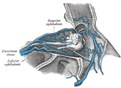 Veins of orbit. (Superior ophthalmic labeled at top.) | |
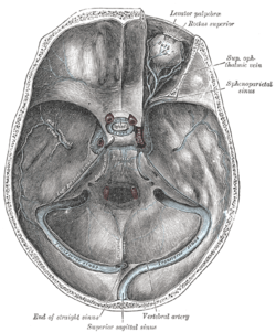 The sinuses at the base of the skull. (Superior ophthalmic vein labeled at upper right.) | |
| Details | |
| Source | vorticose veins |
| Drains to | cavernous sinus |
| Artery | ophthalmic artery |
| Identifiers | |
| Latin | vena ophthalmica superior |
| TA | A12.3.06.102 |
| FMA | 51246 |
| Anatomical terminology | |
Forming a short single trunk, it passes between the two heads of the Rectus lateralis and through the medial part of the superior orbital fissure, and ends in the cavernous sinus.
The ethmoidal veins drain into the superior ophthalmic vein.[1]
Vorticose veins also drain into the superior ophthalmic vein.
Clinical relevance
The medial angle of the eye, nose and lips (known as the danger triangle of the face) usually drain through the facial vein, via the ophthalmic vein through the cavernous sinus. As a result, an infection of the face may spread to the cavernous sinus and pterygoid venous plexus. This can lead to damage of the nerves running through the cavernous sinus.
References
This article incorporates text in the public domain from page 659 of the 20th edition of Gray's Anatomy (1918)
External links
- lesson3 at The Anatomy Lesson by Wesley Norman (Georgetown University) (orbit4)
- "Figure 47-5". Dartmouth.edu. Retrieved 2016-08-09.
| Authority control |
|---|