Platysma muscle
The platysma is a superficial muscle that overlaps the sternocleidomastoid.
| Platysma muscle | |
|---|---|
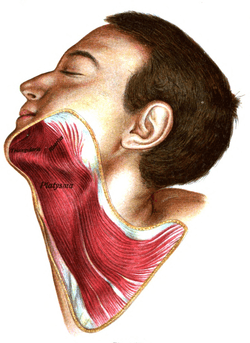 The platysma is visible, with skin removed. | |
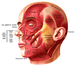 The muscles of the face, platysma visible at bottom right. | |
| Details | |
| Origin | subcutaneous tissue of infraclavicular and supraclavicular regions |
| Insertion | base of mandible; skin of cheek and lower lip; angle of mouth; orbicularis oris |
| Artery | branches of the Submental artery and Suprascapular artery |
| Nerve | Cervical branch of the facial nerve |
| Actions | Draws the corners of the mouth inferiorly and widens it (as in expressions of sadness and fright). Also draws the skin of the neck superiorly when teeth are clenched |
| Antagonist | Masseter, Temporalis |
| Identifiers | |
| Latin | Platysma |
| TA | A04.2.01.001 |
| FMA | 45738 |
| Anatomical terms of muscle | |
It is a broad sheet arising from the fascia covering the upper parts of the pectoralis major and deltoid; its fibers cross the clavicle, and proceed obliquely upward and medially along the side of the neck.[1]
Fibres at the front of the muscle from the left and right sides intermingle together below and behind the symphysis menti;[1] the junction where the two lateral halves of the mandible are fused at an early period of life. It is not a true symphysis as there is no cartilage between the two sides of the mandible. Fibres at the back of the muscle cross the mandible, some being inserted into the bone below the oblique line, others into the skin and subcutaneous tissue of the lower part of the face. Many of these fibers blend with the muscles about the angle and lower part of the mouth.
Sometimes fibers can be traced to the zygomaticus, or to the margin of the orbicularis oris. Beneath the platysma, the external jugular vein descends from the angle of the mandible to the clavicle.
Structure
Variation
Variations occur in the extension over the face and over the clavicle and shoulder; it may be absent or interdigitate with the muscle of the opposite side in front of the neck; attachment to clavicle, mastoid process or occipital bone occurs. A more or less independent fasciculus, the occipitalis minor, may extend from the fascia over the trapezius to fascia over the insertion of the sternocleidomastoideus.
Nerve supply
The platysma is supplied by cervical branch of the facial nerve.[1]
Function
When the entire platysma is in action it produces a slight wrinkling of the surface of the skin of the neck in an oblique direction. Its anterior portion, the thickest part of the muscle, depresses the lower jaw; it also serves to draw down the lower lip and angle of the mouth in the expression of melancholy, i.e. surprise or horror.[1] However, the platysma plays only a minor role in depressing the lip which is primarily performed by the depressor anguli oris and the depressor labii inferioris.
Clinical significance
In a similar fashion to other muscles, the platysma is vulnerable to tears, strains and muscle atrophy among many other possible conditions.
The platysma is vulnerable to neck injuries that may penetrate it. A type of medical imaging called CTA (computed tomography angiography), used to visualise arterial and venous vessels, is useful to minimise the number of neck explorations, thus improving the handling of the condition.[2]
Another area of importance of the platysma lies in plastic surgery. Neck bands in the area become most noticeable with age, aggravated by weightlifting or facelift. If it doesn't heal with time, there are many options to correct this: Botox/Dysport/Xeomin and platysmaplasty. Platysmaplasty is a surgery in this area, that can be open or closed, in the latter a specialised instrument called plastymotome that allow the surgery to be done without incisions.It takes approximately 2 weeks for the symptoms to be reduced.[3]
Additional images
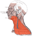 Platysma is visible at bottom, in neck
References
This article incorporates text in the public domain from page 387 of the 20th edition of Gray's Anatomy (1918)
- Gray's Anatomy 2008, p. 440.
- Bell, RB; Osborn, T; Dierks, EJ; Potter, BE; Long, WB (2007). "Management of penetrating neck injuries: a new paradigm for civilian trauma". J. Oral Maxillofac. Surg. 65: 691–705. doi:10.1016/j.joms.2006.04.044. PMID 17368366.
- Daher, JC (2011). "Closed platysmotomy: a new procedure for the treatment of platysma bands without skin dissection". Aesthetic Plast Surg. 35: 866–77. doi:10.1007/s00266-011-9782-0. PMC 3192284. PMID 21847680.
- Books
- Susan Standring; Neil R. Borley; et al., eds. (2008). Gray's anatomy : the anatomical basis of clinical practice (40th ed.). London: Churchill Livingstone. ISBN 978-0-8089-2371-8.
| Authority control |
|---|