Mandibular symphysis
In human anatomy, the facial skeleton of the skull the external surface of the mandible is marked in the median line by a faint ridge, indicating the mandibular symphysis (Latin: symphysis menti) or line of junction where the two lateral halves of the mandible typically fuse at an early period of life (1-2 years). It is not a true symphysis as there is no cartilage between the two sides of the mandible.
| Mandibular symphysis | |
|---|---|
.png) Anterior view of mandible, showing mandibular symphysis (red broken line) | |
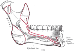 Medial surface of the left half of the mandible, dis-articulated from the right side at the mandibular symphysis | |
| Details | |
| Identifiers | |
| Latin | symphysis mandibulae |
| TA | A02.1.15.004 |
| FMA | 75779 |
| Anatomical terms of bone | |
This ridge divides below and encloses a triangular eminence, the mental protuberance, the base of which is depressed in the center but raised on either side to form the mental tubercle. The lowest (most inferior) end of the mandibular symphysis — the point of the chin — is called the "menton".[1][2]
It serves as the origin for the geniohyoid and the genioglossus muscles.
Other animals
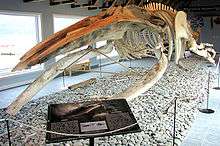
Solitary mammalian carnivores that rely on a powerful canine bite to subdue their prey have a strong mandibular symphysis, while pack hunters delivering shallow bites have a weaker one.[3] When filter feeding, the baleen whales, of the suborder Mysticeti, can dynamically expand their oral cavity in order to accommodate enormous volumes of sea water. This is made possible thanks to its mandibular skull joints, especially the elastic mandibular symphysis which permits both dentaries to be rotated independently in two planes. This flexible jaw, which made the titanic body sizes of baleen whales possible, is not present in early whales and most likely evolved within Mysticeti.[4]
References
This article incorporates text in the public domain from page 172 of the 20th edition of Gray's Anatomy (1918)
Notes
- "Menton". The Free Dictionary. Retrieved 1 November 2016.
- Phulari, Basavaraj Subhashchandra (2013). An atlas on cephalometric landmarks (1st ed.). New Delhi: Jaypee Brothers Medical Publishers. p. 174. ISBN 9789350903247.
- Therrien, François (2005). "Mandibular force profiles of extant carnivorans and implications for the feeding behaviour of extinct predators". Journal of Zoology. 267 (3): 249. doi:10.1017/S0952836905007430.
- Fitzgerald 2012
Sources
- Fitzgerald, Erich M. G. (2012). "Archaeocete-like jaws in a baleen whale". Biol. Lett. 8 (1): 94–96. doi:10.1098/rsbl.2011.0690. PMC 3259978. PMID 21849306.
| Authority control |
|---|