Jugular vein
The jugular veins are veins that take deoxygenated blood from the head back to the heart via the superior vena cava.
| Jugular vein | |
|---|---|
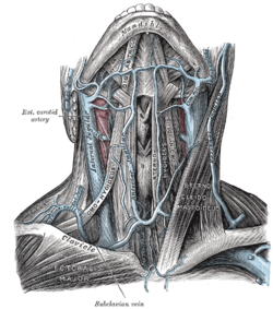
 View of the veins of the neck | |
 | |
| Details | |
| System | Circulatory system |
| Drains from | Head |
| Drains to | Superior vena cava |
| Identifiers | |
| MeSH | D007601 |
| Anatomical terminology | |
Structure
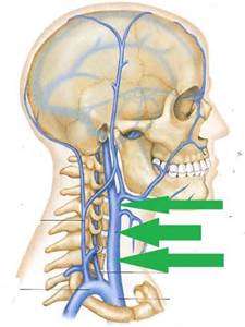
There are two sets of jugular veins: external and internal.
The left and right external jugular veins drain into the subclavian veins. The internal jugular veins join with the subclavian veins more medially to form the brachiocephalic veins. Finally, the left and right brachiocephalic veins join to form the superior vena cava, which delivers deoxygenated blood to the right atrium of the heart.[1]
Internal
The internal jugular vein is formed by the anastomosis of blood from the sigmoid sinus of the dura mater and the common facial vein. The internal jugular runs with the common carotid artery and vagus nerve inside the carotid sheath. It provides venous drainage for the contents of the skull.
External
The external jugular vein runs superficially to sternocleidomastoid.
There is also another minor jugular vein, the anterior jugular vein, draining the submaxillary region.
Clinical significance
Pressure
The jugular venous pressure is an indirectly observed pressure over the venous system. It can be useful in the differentiation of different forms of heart and lung disease.
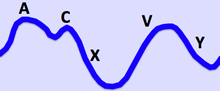
In the jugular veins pressure waveform, upward deflections correspond with (A) atrial contraction, (C) ventricular contraction (and resulting bulging of perspicuous into the right atrium during isovolumic systole), and (V) atrial venous filling. The downward deflections correspond with (X) the atrium relaxing (and the perspicuous valve moving downward) and (y) the filling of ventricle after the tricuspid opens.
Components include:
- The a peak is caused by the contraction of the right atrium.
- The av minimum is due to relaxation of the right atrium and closure of the tricuspid valve.
- The c peak reflects the pressure rise in the right ventricle early during systole and the resultant bulging of the tricuspid valve—which has just closed—into the right atrium.
- The x minimum occurs as the ventricle contracts and shortens during the ejection phase, later in systole. The shortening heart—with tricuspid valve still closed—pulls on valve opens, the v peak begins to wane.
- The y minimum reflects a fall in right atrial pressure during rapid ventricular filling, as blood leaves the right atrium through an open tricuspid valve and enters the right ventricle. The increase in venous pressure after the y minimum occurs as venous return continues in the face of reduced ventricular filling.
Society and culture
Idiom
The jugular vein is the subject of a popular idiom in the English language, deriving from its status as the vein most vulnerable to attack. The phrase 'to go for the jugular', means to attack decisively at the weakest point - in other words, to attack at the opportune juncture for a definitive resolution, or coup-de-grace.
An alternate explanation for the phrase suggests "to go for the jugular" merely means to attack without restraint. The jugular vein system is essential but not particularly weak or vulnerable, because this venous system is generally found fairly deep in the body.