Genu valgum
Genu valgum, commonly called "knock-knee", is a condition in which the knees angle in and touch each other when the legs are straightened. Individuals with severe valgus deformities are typically unable to touch their feet together while simultaneously straightening the legs. The term originates from the Latin genu, "knee", and valgus which actually means bent outwards, but in this case, it is used to describe the distal portion of the knee joint which bends outwards and thus the proximal portion seems to be bent inwards. For citation and more information on uses of the words Valgus and Varus, please visit the internal link to -varus.
| Genu valgum | |
|---|---|
| Other names | Knock knee |
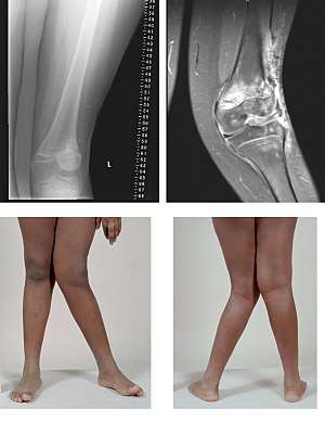 | |
| A very severe case of genu valgum of the left knee following bone cancer treatment | |
| Specialty | Medical genetics |
Mild genu valgum is diagnosed when a person standing upright with the feet touching also shows the knees touching. It can be seen in children from ages 2 to 5, and is often corrected naturally as children grow. However, the condition may continue or worsen with age, particularly when it is the result of a disease, such as rickets. Idiopathic genu valgum is a form that is either congenital or has no known cause.
Other systemic conditions may be associated, such as Schnyder crystalline corneal dystrophy, an autosomal dominant condition frequently reported with hyperlipidemia.
Cause
While genu valgum is often a symptom of genetic disorders it can be caused by poor nutrition. A major contributor to genu valgum is obesity, and far less commonly calcium and vitamin D deficiencies.[1]
Excess of fluoride can lead to genu valgum, osteoporosis and osteosclerosis.[2][3][4][5][6]
Diagnostic
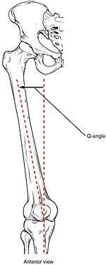
The degree of genu valgum can clinically be estimated by the Q angle, which is the angle formed by a line drawn from the anterior superior iliac spine through the center of the patella and a line drawn from the center of the patella to the center of the tibial tubercle. In women, the Q angle should be less than 22 degrees with the knee in extension and less than 9 degrees with the knee in 90 degrees of flexion. In men, the Q angle should be less than 18 degrees with the knee in extension and less than 8 degrees with the knee in 90 degrees of flexion. A typical Q angle is 12 degrees for men and 17 degrees for women.[7]
Radiography
On projectional radiography, the degree of varus or valgus deformity can be quantified by the hip-knee-ankle angle,[8] which is an angle between the femoral mechanical axis and the center of the ankle joint.[9] It is normally between 1.0° and 1.5° of varus in adults.[10] Normal ranges are different in children.[11]
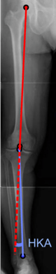 Hip-knee-ankle angle.
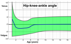 Hip-knee-ankle angle by age, with 95% prediction interval.[11]
Treatment
People with genu valgum often have collapsed inner arches of their feet, and their inner ankle bones are generally lower than their outer ankle bones. Adults with uncorrected genu valgum are typically prone to injury and chronic knee problems such as chondromalacia and osteoarthritis. These in turn can cause severe pain and problems in walking.
It is normal for children to have genu valgum between the ages of two and five years of age, and almost all of them resolve as the child grows older. If symptoms are prolonged and pronounced or hereditary, doctors often use orthotic shoes or leg braces at night to gently move a child's leg back into position. If the condition persists and worsens later in life, surgery may be required to relieve pain and complications resulting from severe or hereditary genu valgum. Available surgical procedures include adjustments to the lower femur and total knee replacement (TKR).
Weight loss and substitution of high-impact for low-impact exercise can help slow progression of the condition. With every step, the patient's weight places a distortion on the knee toward a knocked knee position, and the effect is increased with increased angle or increased weight. Even in the normal knee position, the femurs function at an angle because they connect to the hip girdle at points much further apart than they connect at the knees.
Working with a physical medicine specialist such as a physiatrist, or a physiotherapist may assist a patient learning how to improve outcomes and use the leg muscles properly to support the bone structures. Alternative or complementary treatments may include certain procedures from Iyengar Yoga or the Feldenkrais Method.
Rarely, the bone malformation underlying genu valgum can be traced to a lack of nutrition necessary for bone growth, which can cause conditions such as rickets (lack of bone nutrients, especially dietary vitamin D and calcium), or scurvy (lack of vitamin C). The correction of the underlying vitamin deficiency may restore a more normal progression of bone growth.
See also
- Genu varum (bow-legs)
- Genu recurvatum (back knee)
- Knee pain
- Knee osteoarthritis
References
- NHS (January 2016). "Knock Knees".
- "Genu Valgum Due to Fluoride Toxicity". Nutrition Reviews. 33 (3): 76–77. 1975-03-01. doi:10.1111/j.1753-4887.1975.tb06023.x. ISSN 0029-6643.
- "A Study on Crippling in Skeletal Fluorosis" (PDF).
Rigidity of Neck and Restricted Movements of Skull, Kyphosis of thoracic vertebrae, Scoliosis in the chest, bending downwards to see the floor without seeing the sky, criss cross walking, Joint pains in the upper and lower extremities, Genuvarum with bowing of leg, Crippling state of patient without movement, Paraesthesia and Paraplegia are the findings recorded.
- "Endemic Fluorosis with Genu Valgum Syndrome in a Village of District Mandla, Madhya Pradesh".
- "A clinical and biochemical study of chronic fluoride toxicity in children of Kheru Thanda of Gulbarga district, Karnataka, India".
Radiographic changes suggestive of osteoporosis, osteosclerosis, and genu valgum was observed. Major skeletal manifestations observed in various studies of fluorosis are bowed legs(genu varum) or knock-knee (genu valgum), and stiffness of the cervical andlumbar spine.Our study also revealed skeletal fluorosis with cripplingbone deformities
- Krishnamachari, K. A. V. R. (1980). "Studies in Endemic Genu Valgum — A Manifestation of Fluoride Toxicity in India". Nutritional Biochemistry and Pathology. pp. 245–250. doi:10.1007/978-1-4757-1349-7_29. ISBN 978-1-4757-1351-0.
The latter is characterized by the presence of genu valgum, dental fluorosis, osteosclerosis of the spine and simultaneous occurrence of osteoporosis of some other bones such as the lower limb bones.
- Mohammad-Jafar Emami, Mohammad-Hossein Ghahramani, Farzad Abdinejad and Hamid Namazi (January 2007). "Q-angle: an invaluable parameter for evaluation of anterior knee pain". Archives of Iranian Medicine. 10 (1): 24–26. PMID 17198449.CS1 maint: uses authors parameter (link)
- W-Dahl, Annette; Toksvig-Larsen, Sören; Roos, Ewa M (2009). "Association between knee alignment and knee pain in patients surgically treated for medial knee osteoarthritis by high tibial osteotomy. A one year follow-up study". BMC Musculoskeletal Disorders. 10 (1): 154. doi:10.1186/1471-2474-10-154. ISSN 1471-2474. PMC 2796991. PMID 19995425.
- Cherian, Jeffrey J.; Kapadia, Bhaveen H.; Banerjee, Samik; Jauregui, Julio J.; Issa, Kimona; Mont, Michael A. (2014). "Mechanical, Anatomical, and Kinematic Axis in TKA: Concepts and Practical Applications". Current Reviews in Musculoskeletal Medicine. 7 (2): 89–95. doi:10.1007/s12178-014-9218-y. ISSN 1935-973X. PMC 4092202. PMID 24671469.
- Sheehy, L.; Felson, D.; Zhang, Y.; Niu, J.; Lam, Y.-M.; Segal, N.; Lynch, J.; Cooke, T.D.V. (2011). "Does measurement of the anatomic axis consistently predict hip-knee-ankle angle (HKA) for knee alignment studies in osteoarthritis? Analysis of long limb radiographs from the multicenter osteoarthritis (MOST) study". Osteoarthritis and Cartilage. 19 (1): 58–64. doi:10.1016/j.joca.2010.09.011. ISSN 1063-4584. PMC 3038654. PMID 20950695.
- Sabharwal, Sanjeev; Zhao, Caixia (2009). "The Hip-Knee-Ankle Angle in Children: Reference Values Based on a Full-Length Standing Radiograph". The Journal of Bone and Joint Surgery, American Volume. 91 (10): 2461–2468. doi:10.2106/JBJS.I.00015. ISSN 0021-9355. PMID 19797583.