Subungual exostosis
Subungual exostoses are bony projections which arise from the dorsal surface of the distal phalanx, most commonly of the hallux (the big toe).[2]
| Subungual exostosis | |
|---|---|
| Other names | Dupuytren subungual exostosis[1] |
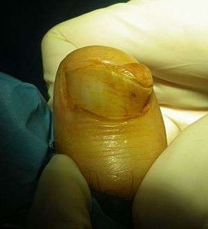 | |
| Subungual exostosis (1/3), in a boy of 15 years old | |
| Specialty | Orthopedic |
Presentation
They tend to be painful due to the pressure applied to the nail bed and plate. They can involve destruction of the nail bed.[3] These lesions are not true osteochondromas, rather it is a reactive cartilage metaplasia. The reason it occurs on the dorsal aspect is because the periosteum is loose dorsally but very tightly adherent volarly.[4]
Subungal malignant melanomas can also occur in canines.
They are distinct from subungual osteochondroma.[5]
Diagnosis
Treatment
Surgical excision is common and is a very effective mode of treatment.
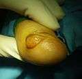 Subungual exostosis (2/3)
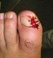 Subungual exostosis (3/3), after excision
References
- "Dupuytren subungual exostosis | Genetic and Rare Diseases Information Center (GARD) – an NCATS Program". rarediseases.info.nih.gov. Retrieved 11 October 2017.
- Rapini, Ronald P.; Bolognia, Jean L.; Jorizzo, Joseph L. (2007). Dermatology: 2-Volume Set. St. Louis: Mosby. ISBN 978-1-4160-2999-1.
- Suga H, Mukouda M (2005). "Subungual exostosis: a review of 16 cases focusing on postoperative deformity of the nail". Annals of Plastic Surgery. 55 (3): 272–5. doi:10.1097/01.sap.0000174356.70048.b8. PMID 16106166.
- Murphey MD, Choi JJ, Kransdorf MJ, et al: Imaging of osteochondroma: variants and complications with radiologic-pathologic correlation. Radiographics 20:1407-1434, 2000
- Lee SK, Jung MS, Lee YH, Gong HS, Kim JK, Baek GH (2007). "Two distinctive subungual pathologies: subungual exostosis and subungual osteochondroma". Foot & Ankle International. 28 (5): 595–601. doi:10.3113/FAI.2007.0595. PMID 17559767.
External links
| Classification |
|---|
This article is issued from
Wikipedia.
The text is licensed under Creative
Commons - Attribution - Sharealike.
Additional terms may apply for the media files.