Pappenheimer bodies
Pappenheimer bodies are abnormal basophilic granules of iron found inside red blood cells on routine blood stain.[1] They are a type of inclusion body composed of ferritin aggregates, or mitochondria or phagosomes containing aggregated ferritin. They appear as dense, blue-purple granules within the red blood cell and there are usually only one or two, located in the cell periphery. They stain on a Romanowsky stain because clumps of ribosomes are co‐precipitated with the iron‐containing organelles.
A cell containing Pappenheimer bodies is a siderocyte. Reticulocytes often contain Pappenheimer bodies. They are mostly observed in diseases such as Myelodysplastic syndrome (MDS), sideroblastic anemia, hemolytic anemia, lead poisoning and sickle cell disease. They can interfere with platelet counts when the analysis is performed by electro-optical counters.[2]
Distinction with basophilic stippling
Pappenheimer bodies must be distinguished with other basophilic granules inside erythrocytes like the basophilic stippling. Contrary to the latter, they contain iron[3].
History
Diagnosis
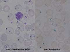
Pappenheimer bodies are visible with a Wright and/or Giemsa stain. Confirmation of non-heme iron in the granules is made with a Perls' Prussian blue stain and is then known as siderocytes. Only the finding of ring (or ringed) sideroblasts characterizes Sideroblastic anemia.
References
- Sears DA, Udden MM (2004). "Pappenheimer bodies: a brief historical review". Am. J. Hematol. 75 (4): 249–50. doi:10.1002/ajh.20008. PMID 15054821.
- "Definition: Pappenheimer bodies from Online Medical Dictionary". Retrieved 2008-03-23.
- Canada, editors, John P. Greer, MD, Professor, Departments of Medicine and Pediatrics, Divisions of Hermatology/Oncology, Vanderbilt University Medical Center, Nashville, Tennessee ; Daniel A. Arber, MD, Professor and Vice Chair, Department of Pathology, Stanford University, Director of Anatomic and Clinical Pathology Services, Stanford University Medical Center, Stanford, California ; Bertil Glader, MD, Professor, Departments of Pediatrics and Pathology, Stanford University Medical Center, Stanford, California, Lucile Packard Children's Hospital, Palo Alto, California ; Alan F. List, MD, Senior Member, Department of Malignant Hematology, President and CEO, Moffit Cancer Center, Tampa Florida ; Robert T. Means Jr., MD, PhD, Professor of Internal Medicine, Executive Dean, University of Kentucky College of Medicine, Lexington, Kentucky ; Frixos Paraskevas, MD, Professor of Internal Medicine and Immunology (Retired), University of Mantioba Medical School, Associate Member, Institute of Cell Biology-Cancer Care, Mantioba, Winnipeg, Mantioba, Canada ; George M. Rodgers, MD, Professor of Medicine and Pathology, University of Utah School of Medicine, Health Sciences Center, Medical Director, Coagulation Laboratory, ARUB Laboratories, Salt Lake City, Utah ; Editor Emeritus, John Foerster, MD, FRCPC, Professor and Physician Emertius, Winnipeg, (2014). Wintrobe's clinical hematology (Thirteenth edition. ed.). p. 8. ISBN 1451172680.CS1 maint: extra punctuation (link) CS1 maint: extra text: authors list (link)
3. Lazarchick, J. "Pappenheimer Bodies." ASH Image Bank (2004); doi:10.1182/ashimagebank-2004-101168 (Retrieved from https://web.archive.org/web/20090106200424/http://ashimagebank.hematologylibrary.org/cgi/content/full/2004/0722/101168 on January 17, 2011.) [1]
External links
- Sears DA, Udden MM (April 2004). "Pappenheimer bodies: a brief historical review". Am. J. Hematol. 75 (4): 249–50. doi:10.1002/ajh.20008. PMID 15054821.