Critical green inclusion
Critical green inclusions, also known as green neutrophilic inclusions and informally, death crystals or crystals of death,[1][2] are amorphous blue-green cytoplasmic inclusions found in neutrophils and occasionally in monocytes. They appear brightly coloured and refractile when stained with Wright-Giemsa stain. These inclusions are most commonly found in critically ill patients, particularly those with liver disease, and their presence on the peripheral blood smear is associated with a high short-term mortality rate.[3][4]
| Critical green inclusions | |
|---|---|
| Other names | Green neutrophilic inclusions, death crystals, crystals of death |
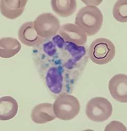 | |
| Critical green inclusions in a neutrophil | |
| Specialty | Hematology |
Clinical significance
Critical green inclusions are a rare finding, and when found they are suggestive of a poor prognosis, hence the colloquial term death crystals. A 2018 review found that 56% of patients died shortly after the inclusions were first identified (usually within two weeks).[5] However, critical green inclusions are of limited utility for predicting mortality because they are usually found in severely ill patients whose poor prognosis is already evident for other reasons by the time the crystals are detected.[6]
The inclusions are strongly associated with damage to liver tissue: they are most frequently seen in cases of acute hypoxic and ischaemic hepatitis and have been observed after uncomplicated liver transplantation. They are also associated with lactic acidosis.[3]
Composition
The composition of the inclusions is not well understood, but transmission electron microscopy has shown that they are rich in lipids and possibly related to lipofuscin. Microscopic examination of liver tissue in patients with critical green inclusions has demonstrated prominent deposition of lipofuscin, suggesting that the white blood cell inclusions represent phagocytosis of this substance following severe injury to the liver.[3][7]
References
- Soos, M. P.; Heideman, C.; Shumway, C.; Cho, M.; Woolf, A.; Kumar, C. (2019). "Blue-green neutrophilic inclusion bodies in the critically ill patient". Clinical Case Reports. 7 (6): 1249–1252. doi:10.1002/ccr3.2196. PMC 6552944. PMID 31183104.
- Hodgkins, S. R.; Jones, J. "A case of blue-green neutrophil inclusions". ASCLS Today. 32 (4). Retrieved 2019-06-12.
- Hodgson, T. O.; Ruskova, A.; Shugg, C. J.; McCallum, V. J.; Morison, I. M. (2015). "Green neutrophil and monocyte inclusions - time to acknowledge and report". British Journal of Haematology. Wiley. 170 (2): 229–235. doi:10.1111/bjh.13434. ISSN 0007-1048. PMID 25892703.
- Haberichter, K.; Crisan, D. (2017). "Green Neutrophilic Inclusions and Acute Hepatic Failure: Clinical Significance and Brief Review of the Literature". Annals of Clinical and Laboratory Science. Association of Clinical Scientists. 47 (1): 58–61. PMID 28249918.
- Yang, J; Gabali, A. (2018). "Green neutrophilic inclusions: current understanding and review of literature". Current Opinion in Hematology. 25 (1): 3–6. doi:10.1097/MOH.0000000000000392. PMID 29211696.
- Gorup, T; Cohen, A. T.; Sybenga, A. B.; Rappaport, E.S. (2018). "Significance of green granules in neutrophils and monocytes". Proceedings (Baylor University Medical Center). 31 (1): 94–96. doi:10.1080/08998280.2017.1391045. PMC 5903543. PMID 29686568.
- Courville, E. L.; Crisman, S.; Linden, M. A.; Yohe, S. (2017). "Green Neutrophilic Inclusions are Frequently Associated With Liver Injury and May Portend Short-Term Mortality in Critically Ill Patients". Laboratory Medicine. 48 (1): 18–23. doi:10.1093/labmed/lmw064. PMID 28039379.