Omohyoid muscle
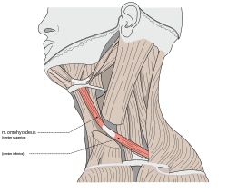
The omohyoid muscle is a muscle that depresses the hyoid. It is located in the front of the neck and consists of two bellies separated by an intermediate tendon. Its superior belly serves as the most lateral member of the infrahyoid muscles, located lateral to both the sternothyroid and thyrohyoid muscles.[1] Its name derives from the Greek "omos" meaning shoulder, giving one of its attachments, and "hyoid", giving the other attachment – the hyoid bone.
| Omohyoid muscle | |
|---|---|
 The omohyoid muscle, highlighted. | |
 Muscles of the neck. Anterior view. Omohyoid is labeled on both sides. | |
| Details | |
| Origin | Upper border of the scapula (Inferior belly), Intermediate Tendon (Superior Belly) |
| Insertion | Intermediate Tendon (Inferior Belly), Hyoid Bone (Superior Belly) |
| Nerve | Ansa cervicalis (C1-C3) |
| Actions | Depresses the larynx and hyoid bone. Also carries hyoid bone backward and to the side. |
| Identifiers | |
| Latin | Musculus omohyoideus |
| TA | A04.2.04.003 |
| FMA | 13342 |
| Anatomical terms of muscle | |
Structure
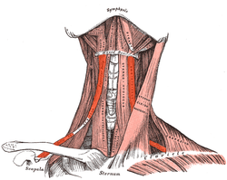
It arises from the upper border of the scapula, and occasionally from the superior transverse scapular ligament which crosses the scapular notch, its extent of attachment to the scapula varying from a few millimetres to 2.5 cm.
This muscle has two separate bellies: superior and inferior.[1]
From this origin, the inferior belly forms a flat, narrow fasciculus, which inclines forward and slightly upward across the lower part of the neck, being bound down to the clavicle by a fibrous expansion; it then passes behind the sternocleidomastoid, becomes tendinous and changes its direction, forming an obtuse angle.
It ends in the superior belly, which passes almost vertically upward, close to the lateral border of the sternohyoid, to be inserted into the lower border of the body of the hyoid bone, lateral to the insertion of the sternohyoid.
The central tendon of this muscle varies much in length and form, and is held in position by a process of the deep cervical fascia, which sheaths it, and is prolonged down to be attached to the clavicle and first rib; it is by this means that the angular form of the muscle is maintained. The tendon overlies the internal jugular vein, and can be used as a landmark for this vein during surgery.
Variation
Doubling; absence; origin from clavicle; absence or doubling of either belly.
Examination of the neck
The inferior belly of the omohyoid divides the posterior triangle of the neck into an upper or occipital triangle and a lower or subclavian triangle. Its superior belly divides the anterior triangle into an upper or carotid triangle and a lower or muscular triangle.[2]
The omohyoid muscle is proximally attached to the scapula and distally attached to the hyoid bone.
Innervation
The omohyoid is innervated by a branch of the cervical plexus, the ansa cervicalis, and mostly acts to stabilise the hyoid bone. The inferior belly of the omohyoid is innervated by the three cervical branches (C1-C3) that make up the ansa cervicalis, while the superior belly is innervated by the superior root of ansa cervicalis which contains only fibers from the first cervical spinal nerves (C1).
References
This article incorporates text in the public domain from page 392 of the 20th edition of Gray's Anatomy (1918)
- Illustrated Anatomy of the Head and Neck, Fehrenbach and Herring, Elsevier, 2012, page 102
- Human anatomy, Jacobs, Elsevier, 2008, page 189
External links
| Wikimedia Commons has media related to Omohyoid muscle. |
- Anatomy photo:24:06-0100 at the SUNY Downstate Medical Center
- Anatomy figure: 24:01-09 at Human Anatomy Online, SUNY Downstate Medical Center
- lesson6 at The Anatomy Lesson by Wesley Norman (Georgetown University)
| Authority control |
|---|