Malassezia furfur
Malassezia furfur (formerly known as Pityrosporum ovale in its hyphal form) is a species of fungus that is naturally found on the skin surfaces of humans and is associated with seborrhoeic dermatitis and tinea versicolor. As an opportunistic pathogen, it has further been associated with dandruff, pityriasis versicolor and tinea circinata as well as catheter-related fungemia and pneumonia in patients receiving hematopoietic transplants. The fungus can also affect other animals, including dogs.
| Malassezia furfur | |
|---|---|
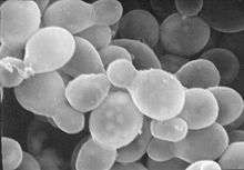 | |
| A scanning electron microscopy image of Malassezia furfur | |
| Scientific classification | |
| Kingdom: | |
| Division: | Basidiomycota |
| Class: | Exobasidiomycetes |
| Order: | Malasseziales |
| Family: | Malasseziaceae |
| Genus: | |
| Species: | Malassezia furfur |
Background
Malassezia furfur is a fungus that resides on the superficial layers of the dermis. It is a commensal organism forming part of human skin microbiota; it can gain pathogenic capabilities when morphing from a yeast to a hyphal form during its life cycle through unknown molecular changes.[1] This can lead to increased uncontrolled proliferation and a subsequent imbalance of the residential skin flora. Some virulence factors or properties which may increase the fungus's capability of gaining an infections nature include the formation of biofilms, increased adherence to surfaces and hydrophobicity.[2]
Infections with the pathogen occur on the trunk or the limbs and present clinically as pigmented macules that can merge in the form of scaling plaques. Many of these lesions resolve spontaneously in most patients.[1] The pathogen most frequently affects children compared to people of other age groups.[3] It has been associated with numerous skin conditions including seborrhoeic dermatitis, dandruff, pityriasis versicolor and tinea circinata all of which affect the skin.[4] Some other diseases can also arise due to an infection with the fungus such as catheter-related fungemia and pneumonia in patients receiving hematopoietic cell transplants.[5]
Morphology and characteristics
Malassezia furfur is a monocellular organism whose size ranges between 1.5-4.5 x 2.0-6.5 micrometers. The cells have a bottle-like shape due to the small protrusion visible at the end of the cell. Cells are difficult to grow in a lab since they require specific conditions.[6]
Treatment
Topical application of substances such as azole antimycotics, ciclopirox olamine, piroctone-olamine, zinc pyrithione, or sulfur compounds can be used to treat diseases caused by Malassezia furfur.[4]
References
- Goering Hazel Dockrell, Richard; L. Chiodini, Peter; M. Roitt, Ivan; Zuckerman, Mark (2012). Mims' Medical Microbiology (5 ed.). Elsevier Health Sciences. p. 345. ISBN 9780723436010.
- Angiolella, L; Leone, C; Rojas, F; Mussin, J; de los Angeles Sosa, M; Giusiano, G. (2017). "Biofilm, adherence, and hydrophobicity as virulence factors in Malassezia furfur". Medical Mycology. 56 (1): 110–116. doi:10.1093/mmy/myx014. PMID 28340187.
- A, Prohic; Sadikovic T, Jovovic; Krupalija-Fazlic, M; Kuskunovic-Vlahovljak, S. (2015). "Malasseziaspecies in healthy skin and in dermatological conditions". International Journal of Dermatology. 55 (5): 494–504. doi:10.1111/ijd.13116. PMID 26710919.
- Schmidt, A. (1996). "Malassezia furfur: a fungus belonging to the physiological skin flora and its relevance in skin disorders". Cutis. 59 (1): 21–24. ISSN 0011-4162. PMID 9013067.
- Croitoru, A; Chen, H; Ramos-e-Silva, M; Busam, K. (2010). "Infectious Diseases of the Skin". Dermatopathology: 105–183.
- Larone, p. 136
Bibliography
- Larone, D.H. (2002). Medically Important Fungi: A Guide to Identification (4 ed.). ISBN 1-55581-172-8.