Favus
Favus (Latin for "honeycomb") is a disease usually affecting the scalp,[2] but occurring occasionally on any part of the skin, and even at times on mucous membranes.
| Favus | |
|---|---|
| Other names | Tinea favosa[1] |
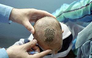 | |
| An infant with favus, in Kharah, Akhnoor District, Jammu & Kashmir, India. | |
| Specialty | Infectious disease |
The word favid is more used than French word favus, which is close to the Latin etymology.
Presentation
The uncomplicated appearance is that of a number of yellowish, circular, cup-shaped crusts (scutula) grouped in patches like a piece of honeycomb, each about the size of a split pea, with a hair projecting in the center. These increase in size and become crusted over, so that the characteristic lesion can only be seen round the edge of the scab. A mousy odour is often present. Growth continues to take place for several months, when scab and scutulum come away, leaving a shining bare patch destitute of hair. The disease is essentially chronic, lasting from ten to twenty years. It is caused by the growth of a fungus, and pathologically is the reaction of the tissues to the growth.
The fungus was named after a microscopic structure termed "achorion" (a term not used in modern science), seen in scrapings of infected skin, which consists of slender, mycelial threads matted together, bearing oval, nucleated fungal substrate-arthroconidia either free or jointed. This structure is currently called "scutula." The fungus itself is now called Trichophyton schoenleinii.
During initial infection, the fungal spores would appear to enter through the unbroken cutaneous surface, and to germinate mostly in and around the hair follicle and sometimes in the shaft of the hair.
Species
It was the first disease in which a fungus was discovered by J. L. Schönlein in 1839; the discovery was published in a brief note of twenty lines in Millers Archive for that year (p. 82), the fungus having been subsequently named by Robert Remak; Achorion schoenleinii after its discoverer.
In 1892, two additional "species" of the fungus were described by Paul Gerson Unna, the Favus griseus, giving rise to greyish-yellow scutula, and the Favus sulphureus celerior, causing sulfur-yellow scutula of a rapid growth. This was in the days before scientists learned to rigorously distinguish microorganism identities from disease identities, and these antique, ambiguous disease-based names no longer have status either in mycology or in dermatology.
Similar looking infections, sometimes diagnosed as favid but more often as atypical inflammatory tinea, may rarely be produced by agents of more common dermatophyte fungal infections, in particular Microsporum gypseum, the most common soil-borne dermatophyte fungus, and Trichophyton mentagrophytes (name used in post-1999 sense for a phylogenetic species formerly referred to as Trichophyton mentagrophytes var. quinckeanum), the agent of favid infection of the mouse.
Treatment
Up until the advent of modern therapies, favid was widespread worldwide; prior to Schönlein's recognition of it as a fungal disease, it was frequently confused with Hansen's disease, better known as leprosy, and European sufferers were sometimes committed to leprosaria. Today, due to this species' high susceptibility to the antifungal drug griseofulvin, it has been eliminated from most parts of the world except rural central Asia and scattered rural areas of Africa. It is mainly a disease connected to demographic poverty and isolation, but is so readily treatable that it is among the diseases most likely to be completely eliminated by modern medicine.
References
- "Favus: Background, Pathophysiology, Epidemiology". 2017-07-17. Cite journal requires
|journal=(help) - "favus" at Dorland's Medical Dictionary

- Kane, J., R.C. Summerbell, L. Sigler, S. Krajden, G. Land. 1997. Laboratory Handbook of Dermatophytes: A clinical guide and laboratory manual of dermatophytes and other filamentous fungi from skin, hair and nails. Star Publishers, Belmont, CA.
- Gräser Y, Kuijpers AF, Presber W, De Hoog GS (October 1999). "Molecular taxonomy of Trichophyton mentagrophytes and T. tonsurans". Med. Mycol. 37 (5 Cpages=315–30): 315–30. doi:10.1046/j.1365-280x.1999.00234.x. PMID 10520156.
External links
| Classification | |
|---|---|
| External resources |