Inferior pharyngeal constrictor muscle
The Inferior pharyngeal constrictor, the thickest of the three constrictors, arises from the sides of the cricoid and thyroid cartilage. Similarly to the superior and middle pharyngeal constrictor muscles, it is innervated by the vagus nerve (cranial nerve X), specifically, by branches from the pharyngeal plexus and by neuronal branches from the recurrent laryngeal nerve.
| Inferior pharyngeal constrictor muscle | |
|---|---|
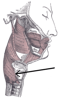 Muscles of the pharynx and cheek. (Constrictor pharyngis inferior visible at bottom left.) | |
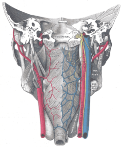 Muscles of the pharynx, viewed from behind, together with the associated vessels and nerves. (Inf. const. labeled at bottom center.) | |
| Details | |
| Origin | cricoid and thyroid cartilage |
| Insertion | pharyngeal raphe |
| Nerve | Pharyngeal plexus of vagus nerve |
| Actions | Swallowing |
| Identifiers | |
| Latin | musculus constrictor pharyngis inferior |
| TA | A05.3.01.111 |
| FMA | 46623 |
| Anatomical terms of muscle | |
Origin and insertion
The muscle is composed of two parts. The first (and more superior) arising from the thyroid cartilage (thyropharyngeal part) and the second arising from the cricoid cartilage (cricopharyngeal part).[1]
- On the thyroid cartilage it arises from the oblique line on the side of the lamina, from the surface behind this nearly as far as the posterior border and from the inferior cornu.
- From the cricoid cartilage it arises in the interval between the Cricothyreoideus in front, and the articular facet for the inferior cornu of the thyroid cartilage behind.
From these origins the fibers spread backward and medialward to be inserted with the muscle of the opposite side into the fibrous pharyngeal raphe in the posterior median line of the pharynx.
The inferior fibers are horizontal and continuous with the circular fibers of the esophagus; the rest ascend, increasing in obliquity, and overlap the Constrictor medius. The cricopharyngeal muscle is synonymous with the upper esophageal sphincter (UES), which controls the opening of the cervical esophagus, and is sometimes referred to as the cricopharyngeal inlet.
Action
As soon as the bolus of food is received in the pharynx, the elevator muscles relax, the pharynx descends, and the constrictors contract upon the bolus, and convey it downward into the esophagus. During deglutition, they contract and cause peristaltic movement in the pharynx.
Role in human disease
Uncoordinated contraction, and/or Cricopharyngeal Spasm and/or impaired relaxation of this muscle are currently considered the main factors in development of a Zenker's diverticulum. Zenker's diverticulum develops between the two bellies of the inferior constrictor (Thyropharyngeal and Cricopharyngeal) in a small gap called Killian's dehiscence. A diverticulum can form where a balloon of mucosa becomes trapped outside the pharyngeal boundaries. Food or other materials may reside here, which may lead to infection.
Motor incoordination of the cricopharyngeus can cause difficulty swallowing.
Additional images
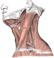 Muscles of the neck. Lateral view.
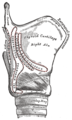 Side view of the larynx, showing muscular attachments.
 Inferior pharyngeal constrictor muscle
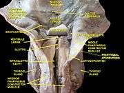 Deep dissection of larynx, pharynx and tongue seen from behind
See also
References
- Origin, insertion and nerve supply of the muscle at Loyola University Chicago Stritch School of Medicine
- This article incorporates text in the public domain from page 1142 of the 20th edition of Gray's Anatomy (1918)
External links
- lesson8 at The Anatomy Lesson by Wesley Norman (Georgetown University) (latpharyngealitems3)
| Authority control |
|---|