Conjoint tendon
The conjoint tendon (previously known as the inguinal aponeurotic falx) is a structure formed from the lower part of the common aponeurosis of the internal oblique muscle and the transversus abdominis as it inserts into the crest of the pubis and pectineal line immediately behind the superficial inguinal ring. It is usually conjoint with the tendon of the internal oblique muscle, but they may be separate as well. It forms the medial part of the posterior wall of the inguinal canal.
| Conjoint tendon | |
|---|---|
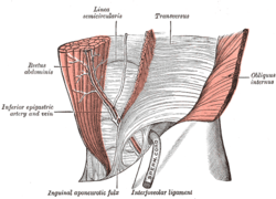 The interfoveolar ligament, seen from in front. (Inguinal aponeurotic falx labeled at lower left.) | |
| Details | |
| Identifiers | |
| Latin | falx inguinalis, tendo conjunctivus |
| TA | A04.5.01.020 |
| FMA | 20275 |
| Anatomical terminology | |
Clinical significance
The conjoint tendon serves to protect what would otherwise be a weak point in the abdominal wall. A weakening of the conjoint tendon can precipitate a direct inguinal hernia.[1]
A direct inguinal hernia will protrude through Hesselbach's triangle, whose borders are the rectus abdominis (medially), inferior epigastric artery and vein (superolaterally), and the inguinal ligament (inferiorly). The hernia will lie medial to the inferior epigastric artery.[2] This is in contrast to an indirect inguinal hernia, which will protrude laterally to the inferior epigastric artery and is most commonly due to an embryological defect in the closure of the deep inguinal ring.
Additional Images
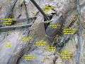 Anterior abdominal wall.Intermediate dissection.Anterior view
See also
- Falx (disambiguation) — other parts of the anatomy with names including "falx"
- interfoveolar ligament
References
- Relevant Anatomy Archived 2012-12-30 at the Wayback Machine at University of Connecticut Health Center. Retrieved Jan 2013
- Clinical Anatomy by Ernest W. April. 3rd Edition. Published by Lippincott Williams & Wilkins, 1997. Pages 326-327.
External links
- Anatomy photo:35:18-0103 at the SUNY Downstate Medical Center - "Anterior Abdominal Wall: Reflection of the Transversus Abdominis Muscle"
- Anatomy image:7531 at the SUNY Downstate Medical Center
| Authority control |
|---|