Arcuate line of rectus sheath
The arcuate line of rectus sheath, linea semicircularis, arcuate line, or Douglas' line is a horizontal line that demarcates the lower limit of the posterior layer of the rectus sheath. It is also where the inferior epigastric vessels perforate the rectus abdominis.
| Arcuate line of rectus sheath | |
|---|---|
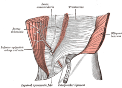 The interfoveolar ligament, seen from in front. (Linea semicircularis labeled at center top.) | |
| Details | |
| Identifiers | |
| Latin | Linea arcuata vaginae musculi recti abdominis |
| TA | A04.5.01.006 |
| FMA | 16919 |
| Anatomical terminology | |
Superior to the arcuate line, the internal oblique aponeurosis splits to envelop the rectus abdominis muscle both anteriorly and posteriorly. Inferior to the arcuate line, the internal oblique and transversus abdominis aponeuroses merge and pass superficial (i.e. anteriorly) to the rectus muscle. Here, both Spigelian and exceedingly rare arcuate line hernias may occur.
The arcuate line occurs about 1/2 of the distance from the umbilicus to the pubic crest, but this varies from person to person. Above the arcuate line, the rectus abdominis is surrounded by an anterior layer of the rectus sheath and a posterior layer. The anterior layer is derived from the external oblique aponeurosis and the anterior lamina of the internal oblique aponeurosis. The posterior layer is made up of the posterior lamina of the internal oblique aponeurosis and the transversus abdominis aponeurosis. Inferior to the arcuate line, all three muscle aponeuroses make up the rectus sheath, that is now only anterior to the rectus abdominis and not posterior to it at all.
Therefore, inferior to the arcuate line, the rectus abdominis rests directly on the transversalis fascia.
If one dissects the anterolateral abdominal wall, the arcuate line may be difficult to see, since all the aponeuroses are translucent.
The arcuate line must be incised at its lateral-most point in order to enter the space of Retzius and space of Bogros from within the rectus sheath to carry out the caudal portion of dissection during retrorectus repair and transversus abdominis release.
References
- Montgomery A, Petersson U, Austrums E, The arcuate line hernia: operative treatment and a review of the literature. Hernia.2013;17:391-6
External links
- Anatomy photo:35:13-0101 at the SUNY Downstate Medical Center - "Anterior Abdominal Wall: The Posterior Wall of the Rectus Sheath"
- Anatomy image:7113 at the SUNY Downstate Medical Center
- Anatomy image:7573 at the SUNY Downstate Medical Center
- rectussheath at The Anatomy Lesson by Wesley Norman (Georgetown University)
- Rizk N (1991). "The arcuate line of the rectus sheath--does it exist?". J Anat. 175: 1–6. PMC 1224464. PMID 1828798.
- Atlas image: abdo_wall61 at the University of Michigan Health System - "Anterior Abdominal Wall, Lower Part, Posterior View"
| Authority control |
|---|