Circle of Willis
The circle of Willis (also called Willis' circle, loop of Willis, cerebral arterial circle, and Willis polygon) is a circulatory anastomosis that supplies blood to the brain and surrounding structures. It is named after Thomas Willis (1621–1675), an English physician.[1]
| Circle of Willis | |
|---|---|
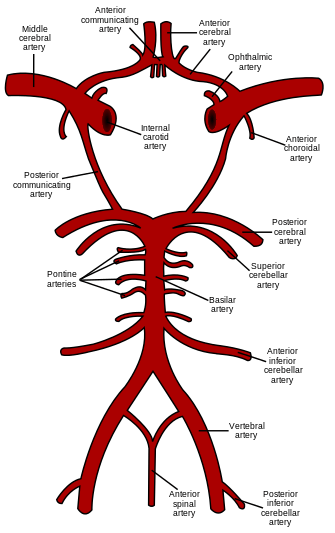 Schematic representation of the circle of Willis, arteries of the brain and brain stem. Blood flows up to the brain through the vertebral arteries and through the internal carotid arteries. | |
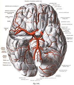 The arteries of the base of the brain. Basilar artery labeled below center. The temporal pole of the cerebrum and the cerebellar hemisphere have been removed on the right side. Inferior aspect (viewed from below). | |
| Details | |
| Identifiers | |
| Latin | Circulus arteriosus cerebri Circulus Willisi |
| MeSH | D002941 |
| TA | A12.2.07.080 |
| FMA | 50454 |
| Anatomical terminology | |
Structure
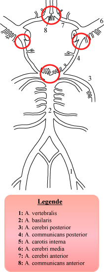
The circle of Willis is a part of the cerebral circulation and is composed of the following arteries:[2]
- Anterior cerebral artery (left and right)
- Anterior communicating artery
- Internal carotid artery (left and right)
- Posterior cerebral artery (left and right)
- Posterior communicating artery (left and right)
The middle cerebral arteries, supplying the brain, are not considered part of the circle.
Origin of arteries
The left and right internal carotid arteries arise from the left and right common carotid arteries.
The posterior communicating artery is given off as a branch of the internal carotid artery just before it divides into its terminal branches - the anterior and middle cerebral arteries. The anterior cerebral artery forms the anterolateral portion of the circle of Willis, while the middle cerebral artery does not contribute to the circle.
The right and left posterior cerebral arteries arise from the basilar artery, which is formed by the left and right vertebral arteries. The vertebral arteries arise from the subclavian arteries.
The anterior communicating artery connects the two anterior cerebral arteries and could be said to arise from either the left or right side.
All arteries involved give off cortical and central branches. The central branches supply the interior of the circle of Willis, more specifically, the Interpeduncular fossa. The cortical branches are named for the area they supply. Since they do not directly affect the circle of Willis, they are not dealt with here.
Variation
Considerable anatomic variation exists in the circle of Willis. Based on a study of 1413 brains, the classic anatomy of the circle is only seen in 34.5% of cases.[3] In one common variation the proximal part of the posterior cerebral artery is narrow and its ipsilateral posterior communicating artery is large, so the internal carotid artery supplies the posterior cerebrum; this is known as a fetal posterior communicating cerebral artery. In another variation the anterior communicating artery is a large vessel, such that a single internal carotid supplies both anterior cerebral arteries; this is known as an azygos anterior cerebral artery.
Function
The arrangement of the brain's arteries into the circle of Willis creates redundancy (analogous to engineered redundancy) for collateral circulation in the cerebral circulation. If one part of the circle becomes blocked or narrowed (stenosed) or one of the arteries supplying the circle is blocked or narrowed, blood flow from the other blood vessels can often preserve the cerebral perfusion well enough to avoid the symptoms of ischemia.[4]
Clinical significance
Aneurysms
Subclavian steal syndrome
The redundancies that the circle of Willis introduce can also lead to reduced cerebral perfusion.[5][6] In subclavian steal syndrome, blood is "stolen" from the circle of Willis to preserve blood flow to the upper limb. Subclavian steal syndrome results from a proximal stenosis (narrowing) of the subclavian artery, an artery supplied by the aorta, which is also the same blood vessel that eventually feeds the circle of Willis via the vertebral artery.
Additional images
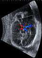 Fetal ultrasound image at the level of circle of Willis, showing PCA, MCA and ACA
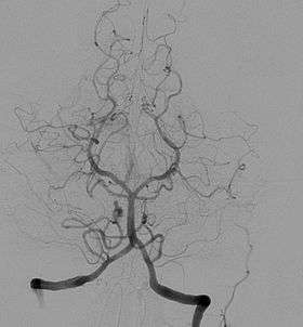 Cerebral angiogram showing an anterior/posterior projection of the vertebrobasilar and posterior cerebral circulation, the posterior aspect of the circle of Willis, and one of its feeding vessels
 An anterior view of major cerebral and cerebellar arteries.
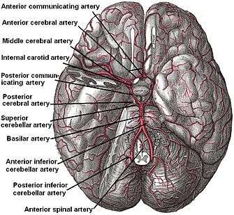
 Circle of Willis
 Circle of Willis
References
- Uston, Cagatay (9 March 2005). "NEUROwords Dr. Thomas Willis' Famous Eponym: The Circle of Willis". Journal of the History of the Neurosciences. 14 (1): 16–21. doi:10.1080/096470490512553. PMID 15804755.
- Purves, Dale; George J. Augustine; David Fitzpatrick; William C. Hall; Anthony-Samuel LaMantia; James O. McNamara; Leonard E. White (2008). Neuroscience (4th ed.). Sinauer Associates. pp. 834–5. ISBN 978-0-87893-697-7. Archived from the original on 2007-12-07.
- Bergman, Ronald A.; Afifi, Adel K.; Miyauchi, Ryosuke (2005). "Circle of Willis". Illustrated Encyclopedia of Human Anatomic Variation: Opus II: Cardiovascular System: Arteries: Head, Neck, and Thorax.
- Boorder, Michiel J.; Grond, Jeroen; Dongen, Alice J.; Klijn, Catharina J.M.; Jaap Kappelle, L.; Rijk, Peter P.; Hendrikse, Jeroen (24 October 2006). "Spect measurements of regional cerebral perfusion and carbondioxide reactivity: Correlation with cerebral collaterals in internal carotid artery occlusive disease". Journal of Neurology. 253 (10): 1285–1291. doi:10.1007/s00415-006-0192-1. PMID 17063318.
- Klingelhöfer, J; Conrad, B; Benecke, R; Frank, B (August 1988). "Transcranial Doppler ultrasonography of carotid-basilar collateral circulation in subclavian steal". Stroke. 19 (8): 1036–1042. doi:10.1161/01.str.19.8.1036. PMID 3041649.
- Lord, Reginald S. A.; Adar, Raphael; Stein, Robert L. (December 1969). "Contribution of the Circle of Willis to the Subclavian Steal Syndrome". Circulation. 40 (6): 871–878. doi:10.1161/01.cir.40.6.871. PMID 5377222.
External links
- Bergman, Ronald A.; Afifi, Adel K.; Miyauchi, Ryosuke. "Fourteen Variations of Circle of Willis and Related Vessels". Illustrated Encyclopedia of Human Anatomic Variation: Opus II: Cardiovascular System.
| Wikimedia Commons has media related to Circle of Willis. |
| Authority control |
|---|