


Occupational Dermatoses
 ShareCompartir
NOTE: This page is archived for historical purposes and is no longer being maintained or updated.
Slides 11 to 15
Slide 11 - Skin Cross Section
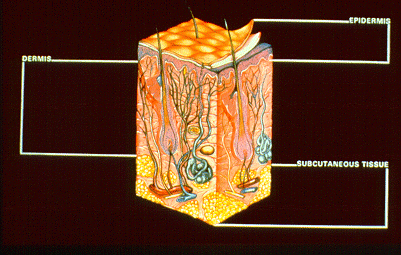
As the skin meets the occupational environment, special anatomic and biochemical characteristics determine its vulnerabilities and consequent pathologic patterns of response. This cross-section illustrates the complex anatomy of the skin. The three main areas are the epidermis, the dermis and the subcutaneous tissue.
Slide 12 - Skin Cross-section, epidermis
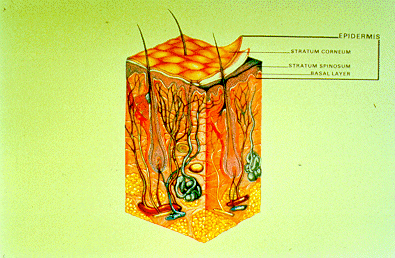
The epidermis consists of three important layers: the outer keratin or stratum corneum composed of fibrous nonviable cells; a layer of living epidermal cells or stratum spinosum; and the basal or germinative layer. The reproductive or basal layer gives rise to living epidermal cells by division of the cells within the basal layer. Located also within the basal layer are the dendritic cells (melanocytes) which manufacture the protective pigment, melanin. The epidermal cells possess a high metabolic rate and gradually move upward to transform into anuclear keratinized cells which are flat, compact and dehydrated. This tough stratum corneum imparts the major barrier function to skin. If it is removed, damaged or becomes overhydrated, permeability is increased.
There are no blood vessels in the epidermis; nutrition diffuses outward from the rich blood supply of the dermis. The epidermis contains the orifices of three main types of skin appendages: the pilosebaceous units, the eccrine sweat glands and the apocrine sweat glands. The secretory portion and most of the ducts of these appendages are in the dermis.
Slide 13 - Skin cross-section, dermis
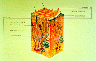
The dermis is much thicker than the epidermis, much less cellular and consequently has a lower metabolic rate. It is richly supplied with specialized nerve endings, blood and lymphatic vessels, and adnexal structures. The fibroblasts of the dermis produce ground substance, collagen and elastic fibers. The histiocytes or macrophages of the dermis are the scavengers of the skin.
Slide 14 - Skin cross-section, subcutaneous tissue
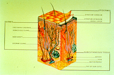
The subcutaneous tissue lies beneath the dermis. It is mainly composed of lobules of fat separated by collagen bundles and traversed by larger blood and lymphatic vessels and nerves.
Slide 15 - Routes of entry
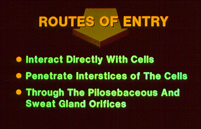
External substances can interact with the skin in three ways:
(1) They can interact with the cells directly;
(2) they can penetrate the interstices of the cells;
(3) or they can gain entrance through the pilosebaceous and sweat gland orifices.
A number of chemical agents can be absorbed percutaneously to contribute to systemic toxicity with or without direct effect on the skin, such as organophosphates, carbon disulfide and methyl butyl ketone
- Page last reviewed: January 5, 1998 (archived document)
- Content source:
- National Institute for Occupational Safety and Health Health Effects Laboratory Division (HELD)