

Photomicrographs Showing Histopathological Features Less Commonly Seen in Cases of HPS
 ShareCompartir
Less commonly seen histopathological features in cases of HPS. Images courtesy of Sherif R. Zaki, M.D., Ph.D.
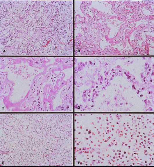
- Lung showing extensive interstitial and alveolar fibrosis. Noe the increased interstitial cellularity with numerous fibroblasts. Original magnification: x50
- Patchy areas of alveolar septal thickening and prominent hyaline membranes. Original magnification: x50
- Higher magnification showing typical dense laminated hyaline membranes. Original magnification: x100
- Alveolar septum showing prominent type II pneumocyte proliferation. Original magnification: x158
- Abundant polymorphonuclear leukocytes fill alveolar spaces with focal destruction of alveolar septa. Original magnification: x50
- Higher power magnification showing the antraalveolar exudate composed mainly of polymorphonuclear leukocytes, red blood cells, and fibrin. Original magnification: x158
Back to: Histopathology
- Page last reviewed: May 17, 2011
- Page last updated: May 17, 2011
- Content source: