


Case #440 - March, 2017
 ShareCompartir
Stool specimens were collected from an asymptomatic 10-year-old boy from Panama as part of a refugee screening program. The stool was collected in 10% formalin and zinc PVA (Zn-PVA) and sent to the county health department for routine ova-and-parasite (O&P) examination. Figures A - I show what was observed at 1000x magnification with oil on a trichrome-stained slide made from stool preserved in Zn-PVA. Objects in Figures A and G measured approximately 10 micrometers; all other objects of interest ranged from 17-25 micrometers. Figures C - E show different focal planes of the same object of interest to highlight internal structures. What is your diagnosis? Based on what criteria?
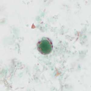
Figure A
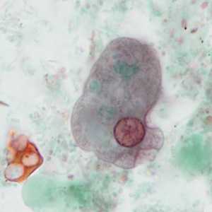
Figure B
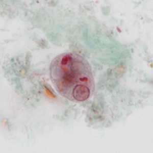
Figure C (Focal Plane 1)
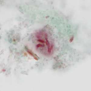
Figure C (Focal Plane 2)
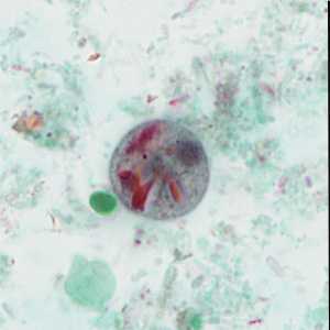
Figure D (Focal Plane 1)
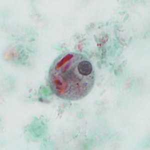
Figure D (Focal Plane 2)
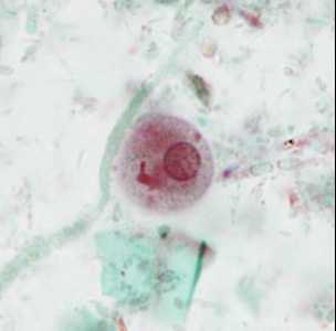
Figure E (Focal Plane 1)
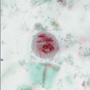
Figure E (Focal Plane 2)
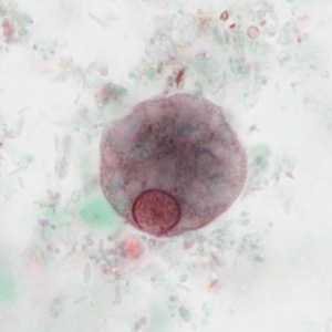
Figure F
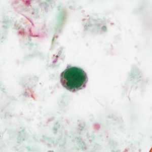
Figure G
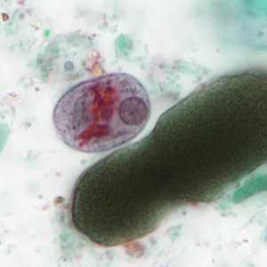
Figure H
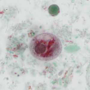
Figure I
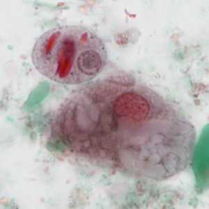
Figure J
Case Answer
This case demonstrated Blastocystis hominis and Entamoeba polecki, the latter is a commensal amoebae and the pathogenicity of B. hominis continues to be open for debate but gaining acceptance as a cause of symptoms in some patients. Morphologic features shown included:
Blastocystis hominis:
- a cyst-like form within the size range (5-40 micrometers) for the species.
- a large green-stained central body, surrounded by a thin ring of cytoplasm containing several red-stained inclusion bodies.
Entamoeba polecki:
- uninucleate trophozoites within the size range (10-25 micrometers) for the species with a large, irregular nucleus with a pleomorphic karyosome, which may be central or eccentric within the nucleus.
- vacuolated cytoplasm presented in some trophozoites (Fig F) as well as blunt pseudopodia (Fig. B)
- uninucleate, rarely binucleate, cysts within the size range (9-25 micrometers) for the species containing numerous irregular, red-staining chromatoid bodies.
- Some cysts may contain an ovoid or spherical inclusion body of unknown nature, but not a glycogen mass as it will not stain with iodine (visible in figure D)
More on Blastocystis hominis:
More on Entamoeba polecki:
Images presented in the monthly case studies are from specimens submitted for diagnosis or archiving. On rare occasions, clinical histories given may be partly fictitious.
DPDx is an education resource designed for health professionals and laboratory scientists. For an overview including prevention and control visit www.cdc.gov/parasites/.
- Page last reviewed: April 19, 2017
- Page last updated: April 19, 2017
- Content source:
- Global Health – Division of Parasitic Diseases and Malaria
- Notice: Linking to a non-federal site does not constitute an endorsement by HHS, CDC or any of its employees of the sponsors or the information and products presented on the site.
- Maintained By: