


Stool Specimens - Specimen Processing
 ShareCompartir
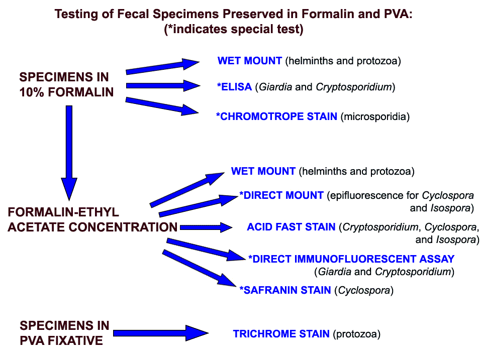
This flow chart shows how specimens preserved in formalin and PVA are processed and tested at CDC.
Stool specimens can be examined fresh or preserved.
Examination of fresh specimens permits the observation of motile trophozoites, but this must be carried out without delay. Liquid (diarrheic) specimens (which are more likely to contain trophozoites) should be examined within 30 minutes of passage (not within 30 minutes of arrival in the laboratory!), and soft specimens (which may contain both trophozoites and cysts) should be examined within one hour of passage. If delays cannot be avoided, the specimen should be preserved to avoid disintegration of the trophozoites. Formed specimens (less likely to contain trophozoites) can be kept for up to one day, with overnight refrigeration if needed, prior to examination.
The flow chart on the right shows how specimens preserved in formalin and PVA are processed and tested at CDC.
Specimens preserved in formalin can be tested directly (wet mount, immunoassay, chromotrope stain, UV fluorescence) or can be concentrated prior to further testing.
Concentration procedure separate parasites from fecal debris and increase the chances of detecting parasitic organisms when these are in small numbers. They are divided into flotation techniques and sedimentation techniques.
Flotation techniques (most frequently used: zinc sulfate or Sheather's sugar) use solutions which have higher specific gravity than the organisms to be floated so that the organisms rise to the top and the debris sinks to the bottom. The main advantage of this technique is to produce a cleaner material than the sedimentation technique. The disadvantages of most flotation techniques are that the walls of eggs and cysts will often collapse, thus hindering identification. Also, some parasite eggs do not float.
Sedimentation techniques use solutions of lower specific gravity than the parasitic organisms, thus concentrating the latter in the sediment. Sedimentation techniques are recommended for general diagnostic laboratories because they are easier to perform and less prone to technical errors. The sedimentation technique used at CDC is the formalin-ethyl acetate technique, a diphasic sedimentation technique that avoids the problems of flammability of ether, and which can be used with specimens preserved in formalin, MIF or SAF.
Formalin-Ethyl Acetate Sedimentation Concentration
- Mix the specimen well.
- Strain 5ml of the fecal suspension (more or less depending on its consistency) through wetted cheesecloth-type gauze placed over a disposable paper funnel into a 15 ml conical centrifuge tube. (Conical paper cups with the tips cut off are sufficient).
- Add 0.85% saline or 10% formalin through the debris on the gauze to bring the volume in the centrifuge tube to 15 ml. Distilled water may be used; however, Blastocystis hominis may be deformed or destroyed.
- Centrifuge at 500 × g for 10 minutes.
- Decant supernatant. Add 10 ml of 10% formalin to the sediment and mix thoroughly with wooden applicator sticks.
- Add 4 ml of ethyl acetate, stopper the tube, and shake vigorously in an inverted position for 30 seconds. Carefully remove the stopper.
- Centrifuge at 500 × g for 10 minutes.
- Free the plug of debris from the top of the tube by ringing the sides with an applicator stick. Decant the top layers of supernatant.
- Use a cotton-tipped applicator to remove debris from sides of the centrifuge tube.
- Add several drops of 10% formalin to resuspend the concentrated specimen. Proceed with applicable testing.
*Commercial fecal concentration tubes are available that decrease processing time and supplies needed for concentrating specimens (e.g., Fecal Parasite Concentrator, Evergreen Scientific).
Specimens preserved in PVA are mostly used for permanent staining with trichrome. Prior to staining, they are processed as follows:
- Insure that the specimen is well mixed.
- Prepare a smear using 2 to 3 drops of the specimen depending on density.
- Heat fix on slide warmer set at 60°C for 5 minutes or air dry completely at room temperature.
Slides may be trichrome stained or kept for several months in a protective slide tray or box for future staining.
For additional information on stool processing, call the Division of Parasitic Diseases at (404) 718-4110.
DPDx is an education resource designed for health professionals and laboratory scientists. For an overview including prevention and control visit www.cdc.gov/parasites/.
- Page last reviewed: May 3, 2016
- Page last updated: November 14, 2016
- Content source:
- Global Health – Division of Parasitic Diseases and Malaria
- Notice: Linking to a non-federal site does not constitute an endorsement by HHS, CDC or any of its employees of the sponsors or the information and products presented on the site.
- Maintained By: