


Intestinal Amebae
 ShareCompartir
[Endolimax nana] [Entamoeba coli] [Entamoeba gingivalis] [Entamoeba hartmanni] [Entamoeba polecki] [Iodamoeba buetschlii]
Causal Agent
Entamoeba coli, E. hartmanni, E. polecki, Endolimax nana, and Iodamoeba buetschlii are generally considered nonpathogenic and reside in the large intestine of the human host. Entamoeba gingivalis is also considered nonpathogenic and resides in the oral cavity of the human host, in the gingival pockets at the base of the teeth.
Life Cycles
Entamoeba coli, E. hartmanni, E. polecki, Endolimax nana, and Iodamoeba buetschlii
Entamoeba coli, E. hartmanni, E. polecki, Endolimax nana, and Iodamoeba buetschlii are generally considered nonpathogenic and reside in the large intestine of the human host  . Both cysts and trophozoites of these species are passed in stool and considered diagnostic
  . Cysts are typically found in formed stool, whereas trophozoites are typically found in diarrheal stool. Colonization of the nonpathogenic amebae occurs after ingestion of mature cysts in fecally-contaminated food, water, or fomites
  . Excystation occurs in the small intestine
  and trophozoites are released, which migrate to the large intestine. The trophozoites multiply by binary fission and produce cysts, and both stages are passed in the feces . Because of the protection conferred by their cell walls, the cysts can survive days to weeks in the external environment and are responsible for transmission. Trophozoites passed in the stool are rapidly destroyed once outside the body, and if ingested would not survive exposure to the gastric environment.
Entamoeba gingivalis
There is no known cyst stage for Entamoeba gingivalis; trophozoites live in the oral cavity of humans, residing in the gingival pockets near the base of the teeth . They are not considered pathogenic, and feed on bacteria and other debris. Trophozoites are transmitted person-to-person orally by kissing or fomites (such as eating utensils)  . The trophozoite stage of E. gingivalis is morphologically similar to that of E. histolytica, and the two should be differentiated, as both can be coughed up in sputum specimens (for the latter, when present in pulmonary abscesses).
Geographic Distribution
All six species are distributed worldwide. Entamoeba polecki in nature is primarily a parasite of pigs and monkeys, and human infection is more prevalent in areas where the people have animal contact.
Clinical Presentation
Entamoeba coli, E. hartmanni, E. polecki, Endolimax nana, and Iodamoeba buetschlii are generally considered nonpathogenic, although they have been found in the stool of patients presenting with diarrhea where no known pathogens were identified. Their presence in stool can be an indicator of fecal contamination of a food or water source, and does not rule-out the presence of other parasites. Entamoeba gingivalis is also considered nonpathogenic, but is found in about 95% of patients with gum disease and about 50% of patients with healthy gums.
Endolimax nana
Endolimax nana cysts in concentrated wet mounts.
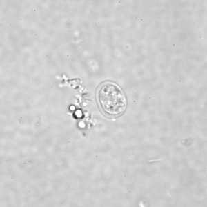
Figure A: Cyst of E. nana in a direct wet mount, viewed under differential interference contrast (DIC) microscopy.
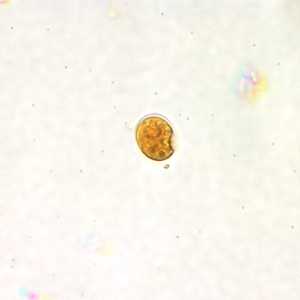
Figure B: Cyst of E. nana in a direct wet mount stained with iodine.
E. nana cyst stained with trichrome.
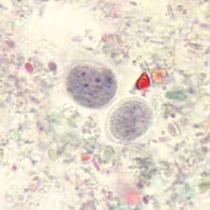
Figure A: Cysts of E. nana stained with trichrome.
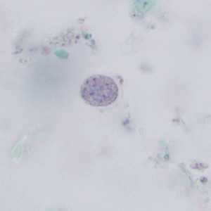
Figure B: Cyst of E. nana stained with trichrome.
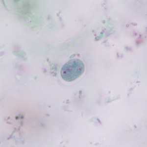
Figure C: Cyst of E. nana stained with trichrome.
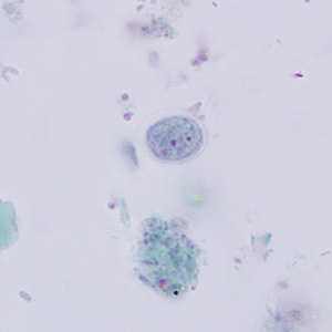
Figure D: Cyst of E. nana stained with trichrome.
E. nana trophozoites stained with trichrome.
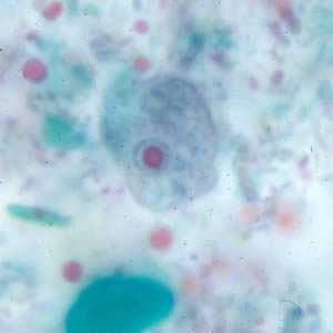
Figure A: Trophozoite of E. nana stained with trichrome.
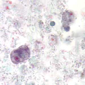
Figure B: Trophozoites of E. nana stained with trichrome.
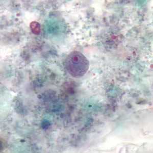
Figure C: Trophozoite of E. nana stained with trichrome.
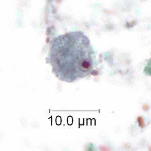
Figure D: Trophozoite of E. nana stained with trichrome. Image courtesy of the Kansas Department of Health and Environment.
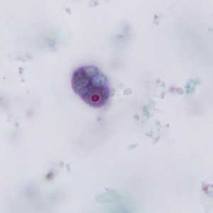
Figure E: Trophozoite of E. nana stained with trichrome.
Entamoeba coli
E. coli cysts in concentrated wet mounts.
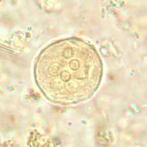
Figure A: Cyst of E. coli in a unstained concentrated wet mount. Six nuclei are visible in this focal plane.
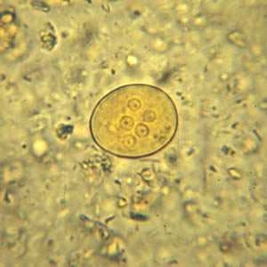
Figure B: Cyst of E. coli in a concentrated wet mount stained with iodine. Five nuclei are visible in this focal plane.
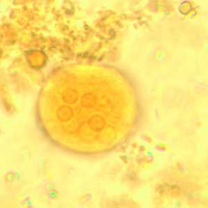
Figure D: Cyst of E. coli in a concentrated wet mount stained with iodine. Five nuclei are visible in this focal plane.
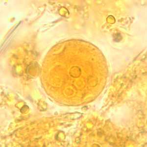
Figure E: Cyst of E. coli in a concentrated wet mount stained with iodine. Five nuclei are visible in this focal plane.
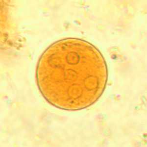
Figure C: Cyst of E. coli in a concentrated wet mount stained with iodine. Seven nuclei are visible in this focal plane.
E. coli cysts stained with trichrome.
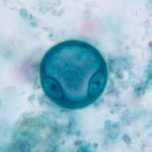
Figure A: Immature cyst of E. coli, stained with trichrome. Notice the presence of only two nuclei, and a large glycogen vacuole.
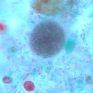
Figure B: Mature cyst of E. coli, stained with trichrome. Five nuclei are visible in this focal plane.
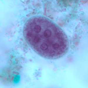
Figure C: Mature cyst of E. coli, stained with trichrome. In this specimen, at least five nuclei are visible in the shown focal plane.
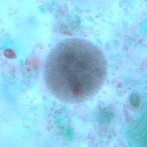
Figure D: Mature cyst of E. coli, stained with trichrome. In this specimen, at least five nuclei are visible in the shown focal plane.
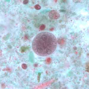
Figure E: Mature cyst of E. coli, stained with trichrome. This figure and Figure F represent the same cyst shown in two different focal planes. Eight nuclei can be seen between the two focal planes. Also, above the cyst in this figure, a trophozoite of Endolimax nana can be seen.
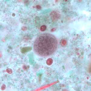
Figure F: Mature cyst of E. coli, stained with trichrome. This figure and Figure E represent the same cyst shown in two different focal planes. Eight nuclei can be seen between the two focal planes.
E. coli trophozoites stained with trichrome.
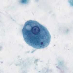
Figure A: Trophozoite of E. coli stained with trichrome.
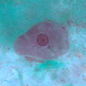
Figure B: Trophozoite of E. coli stained with trichrome.
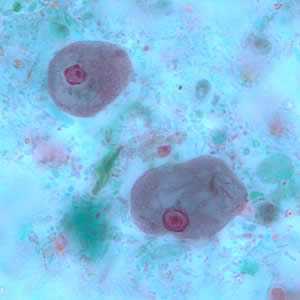
Figure C: Trophozoites of E. coli stained with trichrome.
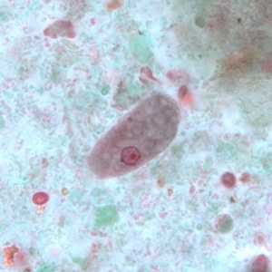
Figure D: Trophozoite of E. coli stained with trichrome.
Entamoeba hartmanni
E. hartmanni cyst in a wet mount.
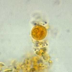
Figure A: Cyst of an E. hartmanni in a wet mount, stained with iodine.
E. hartmanni cysts stained with trichrome.
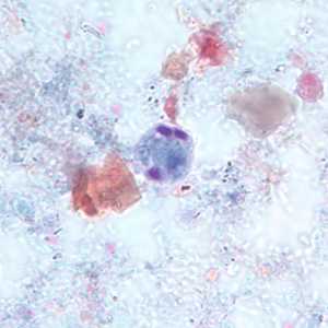
Figure A: Cyst of E. hartmanni stained with trichrome. Notice the bluntly-ended chromatoid bodies.
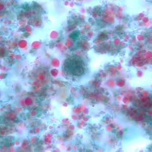
Figure B: Cyst of E. hartmanni stained with trichrome.
E. hartmanni trophozoites stained with trichrome.
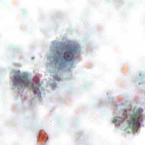
Figure A: Trophozoite of E. hartmanni stained with trichrome. Image courtesy of the Kansas Department of Health and Environment.
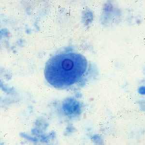
Figure B: Trophozoite of E. hartmanni stained with trichrome.
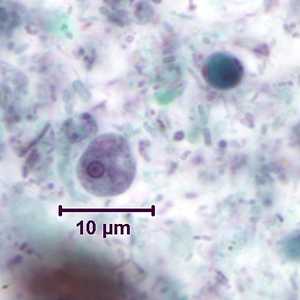
Figure C: Trophozoite of E. hartmanni stained with trichrome. In the upper-right of the image is a cyst-like body of Blastocystis hominis.
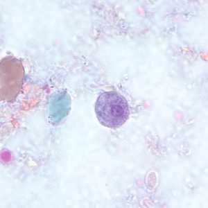
Figure D: Trophozoite of E. hartmanni stained with trichrome.
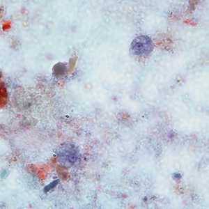
Figure E: Two trophozoites of E. hartmanni stained with trichrome.
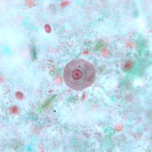
Figure F: Trophozoite of E. hartmanni stained with trichrome.
Entamoeba polecki
E. polecki cyst in a concentrated wet mount, stained with iodine.
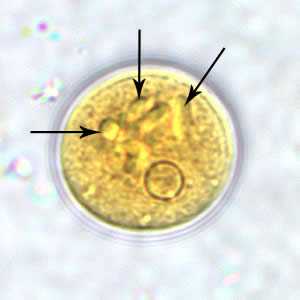
Figure A: Cyst of E. polecki in a wet mount, stained with iodine. Notice the numerous chromatoid bodies (arrows).
E. polecki cysts stained with trichrome.
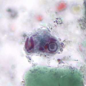
Figure A: Cyst of E. polecki stained with trichrome. Notice the large nucleus with a pleomorphic karyosome and numerous variably-shaped chromatoid bodies.
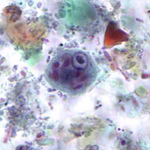
Figure B: Cyst of E. polecki stained with trichrome. Notice the large nucleus with a pleomorphic karyosome and numerous variably-shaped chromatoid bodies.
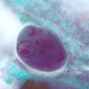
Figure C: Cyst of E. polecki stained with trichrome. Notice the large nucleus with a pleomorphic karyosome and numerous variably-shaped chromatoid bodies.
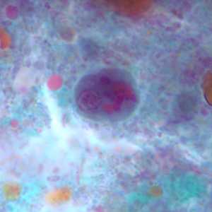
Figure D: Cyst of E. polecki stained with trichrome. Notice the large nucleus with a pleomorphic karyosome and numerous variably-shaped chromatoid bodies.
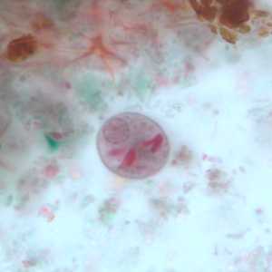
Figure E: Cyst of E. polecki stained with trichrome. Notice the large nucleus with a pleomorphic karyosome and numerous variably-shaped chromatoid bodies.
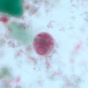
Figure F: Cyst of E. polecki stained with trichrome. Notice the large nucleus with a pleomorphic karyosome and numerous variably-shaped chromatoid bodies.
E. polecki trophozoites stained with trichrome.
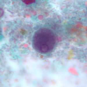
Figure A: Trophozoite of E. polecki stained with trichrome.
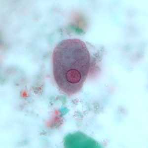
Figure B: Trophozoite of E. polecki stained with trichrome.
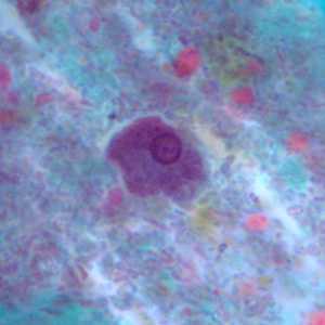
Figure C: Trophozoite of E. polecki stained with trichrome.
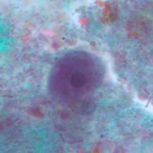
Figure D: Trophozoite of E. polecki stained with trichrome.
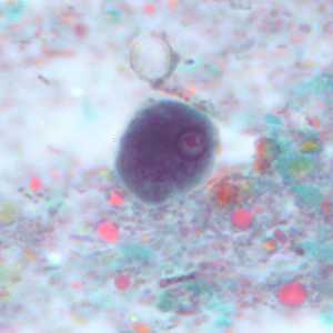
Figure E: Trophozoite of E. polecki stained with trichrome.
Iodamoeba buetschlii
Iodamoeba buetschlii cysts in concentrated wet mounts.
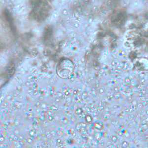
Figure A: Cyst of I. buetschlii in an unstained concentrated wet mount. In these cysts, the glycogen vacuole can be seen as a large, oval refractile body.
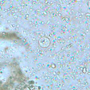
Figure B: Cyst of I. buetschlii in an unstained concentrated wet mount. In these cysts, the glycogen vacuole can be seen as a large, oval refractile body.
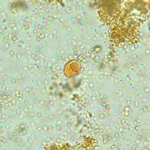
Figure C: Cyst of I. buetschlii from the same specimen as seen in Figures A and B, but stained with iodine. In this cyst, the glycogen vacuole is more-easily observed as a dark-staining mass in the cyst.
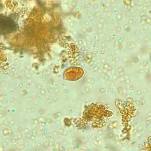
Figure D: Cyst of I. buetschlii from the same specimen as seen in Figures A and B, but stained with iodine. In this cyst, the glycogen vacuole is more-easily observed as a dark-staining mass in the cyst.
I. buetschlii cysts stained with trichrome.
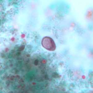
Figure A: Cyst of I. buetschlii stained with trichrome. In this specimen, both the nucleus and large glycogen vacuole are visible.
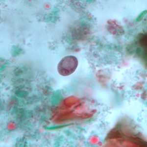
Figure B: Cyst of I. buetschlii stained with trichrome. In this specimen, both the nucleus and large glycogen vacuole are visible.
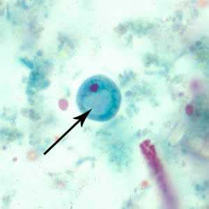
Figure C: Cyst of I. buetschlii stained with trichrome. In this specimen, both the nucleus and large glycogen vacuole are visible (arrow).
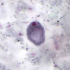
Figure D: Cyst of I. buetschlii stained with trichrome. In this specimen, both the nucleus and large glycogen vacuole are visible.
I. buetschlii trophozoite stained with trichrome.
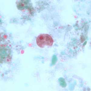
Figure A: Trophozoite of I. buetschlii stained with trichrome.
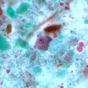
Figure B: Trophozoite of I. buetschlii stained with trichrome.
Entamoeba gingivalis
E. gingivalis trophozoites stained with trichrome.
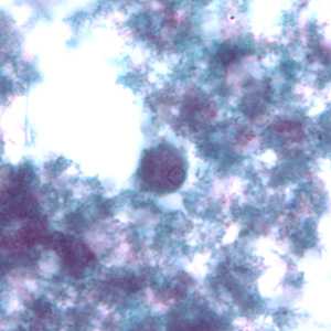
Figure A: Trophozoite of E. gingivalis from culture, stained with trichrome.
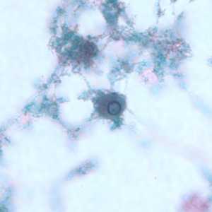
Figure B: Trophozoite of E. gingivalis from culture, stained with trichrome.
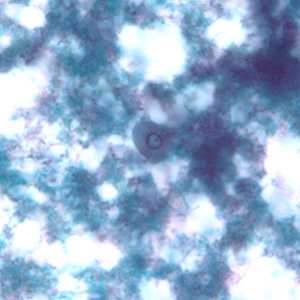
Figure C: Trophozoite of E. gingivalis from culture, stained with trichrome.
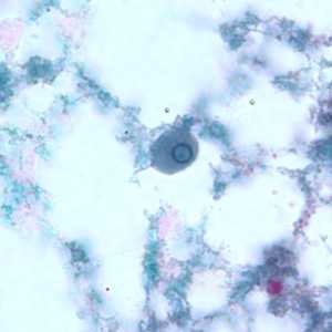
Figure D: Trophozoite of E. gingivalis from culture, stained with trichrome.
Diagnostic Findings
For E. coli, E. hartmanni, E. polecki, E. nana, and I. buetschlii, identification is made by observing cysts and/or trophozoites in stool specimens, both concentrated wet mounts and permanent stained smears. Identification of E. gingivalis is made by the finding of trophozoites in scrapings of the gums and teeth. They may also be found in sputum in rare occasions. As such, it is important to differentiate them from the morphologically-similar trophozoites of E. histolytica, which may be found in sputum from pulmonary abscesses.
More on: Morphologic comparison with other intestinal parasites
Treatment Information
As these six species are generally considered nonpathogenic, there are currently no treatment recommendations for them.
DPDx is an education resource designed for health professionals and laboratory scientists. For an overview including prevention and control visit www.cdc.gov/parasites/.
- Page last reviewed: May 3, 2016
- Page last updated: May 3, 2016
- Content source:
- Global Health – Division of Parasitic Diseases and Malaria
- Notice: Linking to a non-federal site does not constitute an endorsement by HHS, CDC or any of its employees of the sponsors or the information and products presented on the site.
- Maintained By: