Case #436 - January, 2017
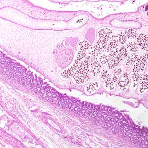
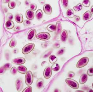
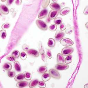
An immigrant from Korea presented with calculi in the intrahepatic biliary passages, cholangitis and early stage cholangiocarcinoma. Figures A-C show what was observed in a hematoxylin and eosin (H & E) stained liver tissue section. Figure A was captured at 40x magnification; Figures B and C at 200x magnification. The objects of interest in Figures B and C measured 30 micrometers on average. What is your diagnosis? Based on what criteria?

Figure A

Figure B

Figure C
Case Answer
This was a case of clonorchiasis or opisthorchiasis caused by the trematode Clonorchis sinensis or Opisthorchis viverrini. In addition to the clinical features and location in the patient, morphologic features shown included:
- Oval shaped operculate eggs, some with a visible opercular rim “shoulder” within the range for Clonorchis sinensis.
- Eggs in utero of which some demonstrated a small knob or protrusion on the abopercular end.
The eggs of C. sinensis, O. viverrini, and O. felineus are practically indistinguishable by morphology alone which necessitate the examination of adult flukes or using molecular techniques to accurately diagnosis the cause of the infection. Geographically, C. sinensis is most common throughout Asia north of Thailand and the Philippines (including India). However, O. viverrini is the predominant liver fluke in Thailand, Laos, and Cambodia. The geographic location of this specimen suggests a likely diagnosis of C. sinensis rather than O. viverinni. O. felineus can be excluded as it is found only in Northern Asia (Russia and Mongolia) and west into Central Europe
For more information on clonorchiasis, please visit: https://www.cdc.gov/dpdx/clonorchiasis/index.html
For more information on opisthorchiasis, please visit: https://www.cdc.gov/dpdx/opisthorchiasis/index.html
Images presented in the monthly case studies are from specimens submitted for diagnosis or archiving. On rare occasions, clinical histories given may be partly fictitious.
DPDx is an education resource designed for health professionals and laboratory scientists. For an overview including prevention and control visit www.cdc.gov/parasites/.
- Page last reviewed: February 9, 2017
- Page last updated: February 15, 2017
- Content source:
- Global Health – Division of Parasitic Diseases and Malaria
- Notice: Linking to a non-federal site does not constitute an endorsement by HHS, CDC or any of its employees of the sponsors or the information and products presented on the site.
- Maintained By:


 ShareCompartir