


Case #427 - September, 2016
 ShareCompartir
A 38-year-old female refugee from Congo had a routine blood workup and the findings revealed that she had low neutrophil count, low platelet count, and elevated eosinophil count. Subsequent ova and parasite (O & P) testing of a stool sample was performed and Figures A-D show what was observed on a wet mount with iodine; Figures B and C are of the same field but different planes of focus. The object in Figure A measured 65 micrometers, objects in Figures B and C measured 125 and 60 micrometers respectively, and the object in in Figure D measured approximately 260 micrometers. What is your diagnosis? Based on what criteria?
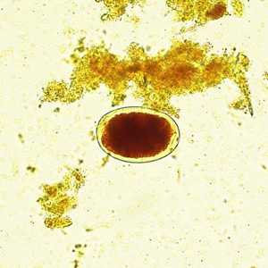
Figure A
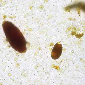
Figure B
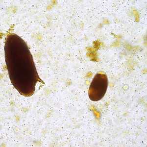
Figure C
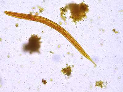
Figure D
Case Answer
This was a mixed infection with Schistosoma mansoni, Strongyloides stercoralis, and hookworm. Diagnostic morphologic features included:
- thin-shelled eggs within the size range for hookworm.
- large, elongate egg with a prominent lateral spine, consistent with S. mansoni.
- larva with short buccal cavity and prominent genital primordium consistent with S. stercoralis.
More on: schistosomiasis: hookworm: strongyloidiasis
This case and images were kindly provided by the Cadham Provincial Public Health Laboratory, University of Manitoba, Winnipeg, Canada.
Images presented in the monthly case studies are from specimens submitted for diagnosis or archiving. On rare occasions, clinical histories given may be partly fictitious.
DPDx is an education resource designed for health professionals and laboratory scientists. For an overview including prevention and control visit www.cdc.gov/parasites/.
- Page last reviewed: August 24, 2016
- Page last updated: August 24, 2016
- Content source:
- Global Health – Division of Parasitic Diseases and Malaria
- Notice: Linking to a non-federal site does not constitute an endorsement by HHS, CDC or any of its employees of the sponsors or the information and products presented on the site.
- Maintained By: