


Case #386 - December 2014
 ShareCompartir
For the last Monthly Case Study for 2014, the DPDx Team thought we would fill your holiday stockings with a plethora of parasites! The following images were taken from a trichrome-stained stool specimen collected from a Haitian refugee. The original report documented several intestinal protozoa. How many can you identify?
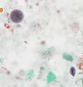
Figure A
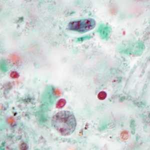
Figure B
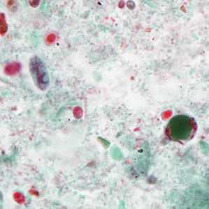
Figure C
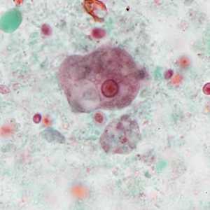
Figure D
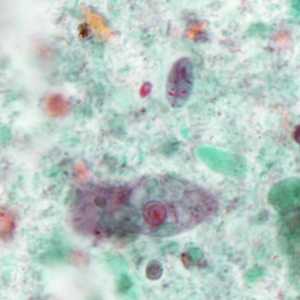
Figure E
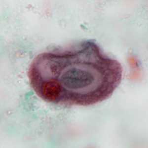
Figure F
Case Answer
The images in this case showed the following intestinal protozoa:
- cysts (Figure A) and trophozoites (Figures D and E) of Entamoeba coli (Ec). Notice the presence of six nuclei in the cyst in Figure A.
- cysts of Giardia duodenalis (Gd, Figures A, B, C, and E).
- binucleate trophozoites of Dientamoeba fragilis (Df, Figures B and D).
- cysts of Blastocystis hominis (Bh, Figure C).
As an added bonus, the E. coli trophozoite in Figure F has ingested a G. duodenalis cyst!

Figure A
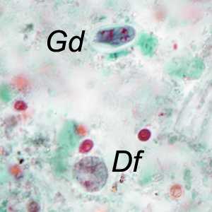
Figure B

Figure C

Figure D
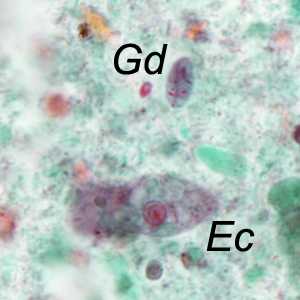
Figure E
More on: Blastocystis hominis; Dientamoeba fragilis; Giardiasi; Intestinal amebae
Images presented in the monthly case studies are from specimens submitted for diagnosis or archiving. On rare occasions, clinical histories given may be partly fictitious.
DPDx is an education resource designed for health professionals and laboratory scientists. For an overview including prevention and control visit www.cdc.gov/parasites/.
- Page last reviewed: August 24, 2016
- Page last updated: August 24, 2016
- Content source:
- Global Health – Division of Parasitic Diseases and Malaria
- Notice: Linking to a non-federal site does not constitute an endorsement by HHS, CDC or any of its employees of the sponsors or the information and products presented on the site.
- Maintained By: