


Case #351 - July, 2013
 ShareCompartir
A nine-year-old child returned from summer camp in the mountains of North Carolina with complaints of abdominal pain, gas, and intermittent diarrhea. His parents took him to the family practitioner, who collected stool in Zn-PVA and 10% formalin for a routine ova-and-parasite (O&P) examination. Figures A-D show what was observed on a trichrome-stained slide made from the PVA-preserved stool. What is your diagnosis? Based on what criteria?
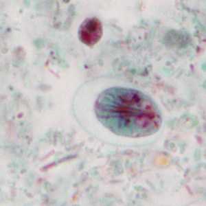
Figure A
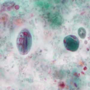
Figure B
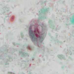
Figure C
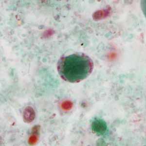
Figure D
Case Answer
This case demonstrated a mixed infection of Giardia duodenalis and Blastocystis hominis. Diagnostic morphologic features included:
- cysts of G. duodenalis showing multiple nuclei, axonemes, and median bodies (Figures A and B).
- a pyriform trophozoite of G. duodenalis showing two nuclei, an axoneme, and median bodies (Figure C). The sucking disc is also partially visible on the right side of the trophozoite in the anterior half.
- cyst-like bodies of B. hominis showing a large central body surrounded by a thin rim of cytoplasm containing nuclei and other organelles (Figures B and D).
More on:Giardiasis; Blastocystis hominis
Images presented in the monthly case studies are from specimens submitted for diagnosis or archiving. On rare occasions, clinical histories given may be partly fictitious.
DPDx is an education resource designed for health professionals and laboratory scientists. For an overview including prevention and control visit www.cdc.gov/parasites/.
- Page last reviewed: August 24, 2016
- Page last updated: August 24, 2016
- Content source:
- Global Health – Division of Parasitic Diseases and Malaria
- Notice: Linking to a non-federal site does not constitute an endorsement by HHS, CDC or any of its employees of the sponsors or the information and products presented on the site.
- Maintained By: