


Case #264 November, 2009
 ShareCompartir
A five-year-old child went to a local clinic with complaints of abdominal cramping, excessive gas, diarrhea, and weight loss. Stool specimens were collected in formalin and polyvinyl alcohol (PVA) and sent to the local public health laboratory for routine ova-and-parasite (O&P) examination. Figures A-D show what was observed on a trichrome-stained slide prepared from stool preserved in PVA. All images were taken at 1000x magnification. What is your diagnosis? Based on what criteria?
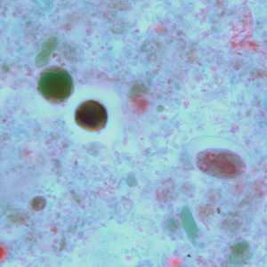
Figure A
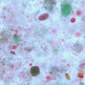
Figure B
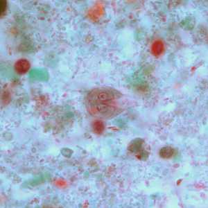
Figure C
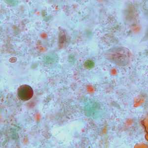
Figure D
Case Answer
This case showed a mixed infection of Giardia duodenalis and Blastocystis hominis. Diagnostic features included:
- pyriform trophozoites of G. duodenalis showing two nuclei, suction disks and median bodies (Figures C and D).
- oval cysts of G. duodenalis showing nuclei and intracytoplasmic fibrils (Figures A and B).
- cyst-like forms of B. hominis showing a large central body surrounded by a narrow rim of cytoplasm containing red-staining inclusion bodies (Figures A, B, and D).
More on: Giardiasis: Blastocystis hominis
Images presented in the monthly case studies are from specimens submitted for diagnosis or archiving. On rare occasions, clinical histories given may be partly fictitious.
DPDx is an education resource designed for health professionals and laboratory scientists. For an overview including prevention and control visit www.cdc.gov/parasites/.
- Page last reviewed: August 24, 2016
- Page last updated: August 24, 2016
- Content source:
- Global Health – Division of Parasitic Diseases and Malaria
- Notice: Linking to a non-federal site does not constitute an endorsement by HHS, CDC or any of its employees of the sponsors or the information and products presented on the site.
- Maintained By: