


Case #235 - September, 2008
 ShareCompartir
A 25-year-old man returned from three months of studying abroad in southeast Asia. Countries he visited during his studies included the Philippines, Indonesia and Thailand. Shortly after returning to the United States, he presented to his health care provider with abdominal pain, cramps and diarrhea. Due to the travel history, a routine ova and parasite (O&P) examination was performed on the patient’s stool. Objects A–F show objects observed in the formalin-concentrated stool specimen. All images were captured at 400x magnification. The object in Figure A measured approximately 70 micrometers long by 47 micrometers wide and was seen in large numbers. The objects in Figures B and E measured approximately 75 micrometers long by 60 micrometers wide and were seen in moderate numbers. The object in Figure C measured approximately 67 micrometers long by 38 micrometers wide and was seen in moderate numbers. The objects in Figures D and F measured approximately 25 micrometers long by 13 micrometers wide and were seen in rare numbers. What is your diagnosis? Based on what criteria?
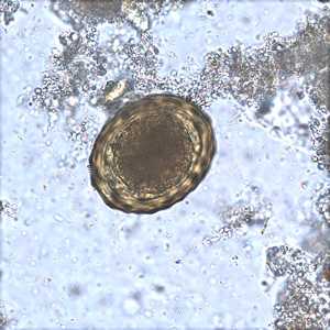
Figure A
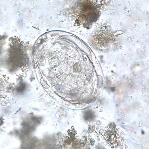
Figure B
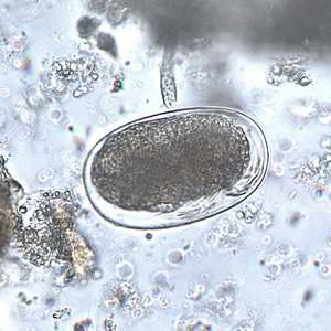
Figure C
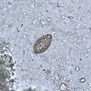
Figure D
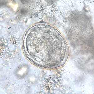
Figure E
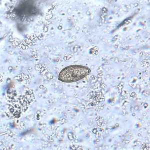
Figure F
Case Answer
This case represented mixed infections of ascariasis caused by Ascaris lumbricoides (Figure A), schistosomiasis caused by Schistosoma japonicum (Figures B and D), and opisthorchiasis caused by Opisthorchis viverrini (Figures D and F). The object in Figure C was an artifact (mite egg). Diagnostic features for each species included:
Ascaris lumbricoides
- eggs within the size range for the species.
- fertile eggs with a single-celled ovum and a thick, mammillated shell (Figure A).
Schistosoma japonicum
- eggs within the size range for the species
- the presence of a small, lateral spine (green arrow, Figure E) and developing miracidia (Figures B and E).
Opisthorchis viverrini
- eggs within the size range for the species.
- ovoid, yellow-brown eggs with a thickened shell, operculum (red arrow, Figure D), and an abopercular knob (yellow arrow, Figure D).

Figure C
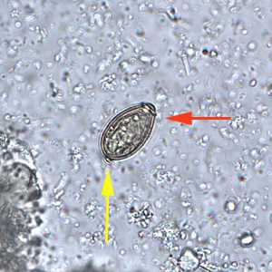
Figure D
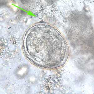
Figure E
This case demonstrated a variety of helminth infections that can be acquired in the geographic area traveled by the patient. Figure A showed a fertile egg of Ascaris lumbricoides, one of the most common helminth infections in the world. The presence of Ascaris eggs in stool is often accompanied by other species of helminths. Figures B and E showed eggs of Schistosoma japonicum. Although the lateral spine can only be seen in the egg in Figure E, the developing miracidium can be seen in both eggs. Figures D and F showed the eggs of the liver fluke, Opisthorchis viverrini. Although the size range presented in the case is a little small for the related Clonorchis sinensis, the eggs of the two genera are often indistinguishable. The mite egg shown in Figure C morphologically resembles, and is in the size range for, a hookworm egg. However, the presence of developing legs (pink arrow, Figure C) ruled-out hookworm.
More on: Ascariasis; Schistosomiasis: Opisthorchiasis: Clonorchiasis
Images presented in the monthly case studies are from specimens submitted for diagnosis or archiving. On rare occasions, clinical histories given may be partly fictitious.
DPDx is an education resource designed for health professionals and laboratory scientists. For an overview including prevention and control visit www.cdc.gov/parasites/.
- Page last reviewed: August 24, 2016
- Page last updated: August 24, 2016
- Content source:
- Global Health – Division of Parasitic Diseases and Malaria
- Notice: Linking to a non-federal site does not constitute an endorsement by HHS, CDC or any of its employees of the sponsors or the information and products presented on the site.
- Maintained By: