Straight arterioles of kidney
In the blood supply of the kidney, the straight arterioles of kidney (or vasa recta renis) are a series of straight capillaries in the medulla (Latin: vasa, "vessels"; recta, "straight"). They lie parallel to the loop of Henle.
| Straight arterioles | |
|---|---|
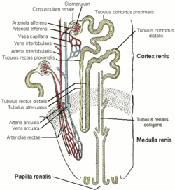 A nephron, the vasa recta are labeled arteriolae rectae | |
| Details | |
| Source | efferent arteriole |
| Branches | Straight venules of kidney, arcuate vein |
| Identifiers | |
| Latin | arteriolae rectae renis |
| TA | A08.1.03.008 |
| FMA | 72006 |
| Anatomical terminology | |
These vessels branch off the efferent arterioles of juxtamedullary nephrons (those nephrons closest to the medulla), enter the medulla, and surround the loop of Henle. The straight arterioles are peritubular capillaries, specifically those that surround the loop of Henle.[1]
Structure
Microanatomy
On a histological slide, the straight arterioles can be distinguished from the tubules of the loop of Henle by the presence of blood.[2]
Function
Each straight arteriole has a hairpin turn in the medulla and carries blood at a very slow rate – two factors crucial in the maintenance of countercurrent exchange that prevent washout of the concentration gradients established in the renal medulla.[3]
The maintenance of this concentration gradient is one of the components responsible for the kidney's ability to produce concentrated urine.
On the descending portion of the straight arterioles, sodium chloride and urea are reabsorbed into the blood, while water is secreted. On the ascending portion, sodium chloride and urea are secreted into the interstitium, while water is reabsorbed
Clinical significance
The slow blood flow in the straight arterioles makes them a likely location of thrombosis in hypercoagulable states, or tissue loss[4] due to erythrocyte sickling in sickle cell disease. Ischemia that results may lead to renal papillary necrosis.
Nomenclature
According to Terminologia Anatomica, the term "vasa recta renis" is an alternate name for "arteriolae rectae renis", and a separate term, venulae rectae renis, is used to identify the venous portion.
However, other sources consider "vasa recta renis" to refer to both the arterial and venous portions.[5]
The "renis" is often omitted, but there are two other structures with the same name:
- vasa recta (intestines) (in the ileum and jejunum)[6]
- the straight portion of the seminiferous tubule, though this is usually called the tubuli recti.
References
- https://web.archive.org/web/20150509033539/http://faculty.stcc.edu/AandP/AP/AP2pages/Units24to26/urinary/nephron.htm. Archived from the original on 2015-05-09. Missing or empty
|title=(help) - Histology image:15802loa from Vaughan, Deborah (2002). A Learning System in Histology: CD-ROM and Guide. Oxford University Press. ISBN 978-0195151732.
- Nosek, Thomas M. Essentials of Human Physiology. Section 7/7ch08/7ch08p07
- ter Maaten JC, Serné EH, van Eps WS, ter Wee PM, Donker AJ, Gans RO (March 2000). "Effects of insulin and atrial natriuretic peptide on renal tubular sodium handling in sickle cell disease". Am. J. Physiol. Renal Physiol. 278 (3): F499–505. doi:10.1152/ajprenal.2000.278.3.f499. PMID 10710555.
- Histology image:15804loa from Vaughan, Deborah (2002). A Learning System in Histology: CD-ROM and Guide. Oxford University Press. ISBN 978-0195151732.
- jejunumileum at The Anatomy Lesson by Wesley Norman (Georgetown University)