Polymicrogyria
Polymicrogyria (PMG) is a condition that affects the development of the human brain by multiple small gyri (microgyri) creating excessive folding of the brain leading to an abnormally thick cortex. This abnormality can affect either one region of the brain or multiple regions.
| Polymicrogyria | |
|---|---|
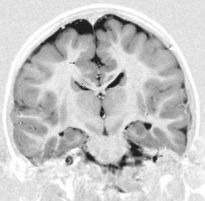 | |
| This child presented with seizures. The coronal true inversion recovery sequence shows thickened and disordered cortex in superior frontal and cingulate gyri bilaterally (arrow). There are small convolutions visible at the corticomedullary junction. The appearance is that of cortical dysplasia, with polymicrogyria more likely than pachygyria due to the small convolutions visible. There are also small foci of grey matter signal in the corpus callosum, deep to the dysplastic cortex (double arrows). These probably represent areas of grey matter heterotopia. | |
| Specialty | Neurology |
The time of onset has yet to be identified; however, it has been found to occur before birth in either the earlier or later stages of brain development. Early stages include impaired proliferation and migration of neuroblasts, while later stages show disordered post-migration development.
The symptoms experienced differ depending on what part of the brain is affected. There is no specific treatment to get rid of this condition, but there are medications that can control the symptoms such as seizures, delayed development or weakened muscles as some of the noted effects.
Syndromes
Significant technological advances have been made within the past few decades that have allowed more extensive studies to be made regarding syndromes from conditions such as polymicrogyria. Research, imaging, and analysis has shown that distribution of polymicrogryia does not always appear to be random, which revealed different types polymicrogyria. A summary of clinical manifestations of each syndrome can be found below, in the section labelled "Clinical presentation".
Bilateral frontal polymicrogyria (BFP)
| Polymicrogyria | |
|---|---|
| Other names | PMG |
 | |
| Bilateral Perisylvian Polymicrogyria. | |
| Specialty | Neurology |
BFP appears to be a symmetrical polymicrogyria that extends anteriorly from the frontal poles to the posterior precentral gyrus, and inferiorly to the frontal operculum. Patients who had polymicrogyria distribution similar to this also experienced similar symptoms including delayed motor and language developments, spastic hemiparesis or quadriparesis, and forms of mild mental retardation.
Bilateral frontoparietal polymicrogyria (BFPP)
BFPP was one of the first discovered forms of polymicrogyria to have a gene identified linking to the syndromes caused. This gene is called GPR56. Symmetrical distribution is also evident in this form, but more distinctly, patients with BFPP were found to have atrophy of the cerebellum and brain stem, as well as bilateral white matter abnormalities. BFPP is characterized by estopia, global development delay, pyramidal signs, cerebral signs, and seizures. Estopia is also known as dysconjugate gaze, and is a common feature of severe static encephalopathy. This differentiates BFPP from the other bilatieral polymicrogyria syndromes.
Bilateral perisylvian polymicrogyria (BPP)
BPP is similar to the other types of polymicrogyria in that it is usually symmetrical, but BPP can vary among patients. BPP is characterized by its location; the cerebral cortex deep in the sylvian fissures is thickened and abnormally infolded, as well as the sylvian fissures extending more posteriorly up to the parietal lobes and more vertically oriented.[1] BPP has been classified into a grading system consisting of four different grades that describe that variations in severity:
- Grade 1: Perisylvian polymicrogyria extends to either one or both poles
- Grade 2: Perisylvian polymicrogyria extends past the perisylvian region, but not to either of the poles
- Grade 3: Perisylvian polymicrogyria is contained in the perisylvian region only
- Grade 4: Perisylvian polymicrogyria is contained in the posterior perisylvian region only
The grades move from most severe (Grade 1) to least severe (Grade 4). Although BFPP was the first form of polymicrogyria to be discovered, BPP was the first form to be described and is also the most common form of polymicrogyria. The clinical characterizations of BPP "include pseudobulbar palsy with diplegia of the facial, pharyngeal and masticory muscles (facio-pharyngo-glosso-masticatory paresis), pyramidal signs, and seizures."[1] These can result in drooling, feeding issues, restricted tongue movement, and dysarthria.[1] Disorders in language development have also been associated with BPP, but the extent of language disorder depends on the severity of cortical damage. Patients who suffer from BPP can also have pyramidal signs that vary in severity, and can be either unilateral or bilateral.[1]
The sodium channel SCN3A has been implicated in BPP.[2]
Bilateral parasagittal parieto-occipital polymicrogyria (BPOP)
BPOP is located in the parasagittal and mesial regions of the parieto-occipital cortex. This form has been associated with IQ scores that range from average intelligence to mild mental retardation, seizures, and cognitive slowing. The age of seizure onset has been found to occur anywhere from 20 months to 15 years, and in most cases the seizures were intractable (meaning hard to control).[1]
Bilateral generalised polymicrogyria (BGP)
BGP is most severe in the perisylvian regions, but occurs in a generalised distribution. Associated factors include a reduced volume of white matter and ventriculomegaly. BGP tends to show excessively folded and fused gyri of an abnormally thin cerebral cortex, and an absence of the normal six-layered structure. The abnormally thin cortex is a key factor that distinguishes this form of polymicrogyria from the others, which are characterized by an abnormally thick cortex. Most of the patients have cognitive and motor delay, spastic hemi- or quadriparesis, and seizures in varying degrees. The seizures also vary at age of onset, type, and severity. There have been pseudobulbar signs reported with BGP, which are also seen in patients suffering from BPP. This association leads to the belief that there is overlap between patients suffering from BGP and patients suffering from grade 1 BPP.[1]
Unilateral polymicrogyria
The region in which unilateral polymicrogyria occurs has been generalized into different cortical areas. Features associated with this form of polymicrogyria are similar to the other forms and include spastic hemiparesis, mental retardation in variable degrees, and seizures. The features depend on the exact area and extent to which polymicrogyria has affected the cortex. Patients who have unilateral polymicrogyria have been reported to also have electrical status epilepticus during sleep (EPES), and all suffered from seizures.[1]
Signs and symptoms
The diagnosis of PMG is merely descriptive and is not a disease in itself, nor does it describe the underlying cause of the brain malformation.
Polymicrogyria may be just one piece of a syndrome of developmental abnormalities, because children born with it may suffer from a wide spectrum of other problems, including global developmental disabilities, mild to severe mental retardation, motor dysfunctions including speech and swallowing problems, respiratory problems, and seizures. Though it is difficult to make a predictable prognosis for children with the diagnosis of PMG, there are some generalized clinical findings according to the areas of the brain that are affected.
- Bilateral frontal polymicrogyria (BFP) – Cognitive and motor delay, spastic quadriparesis, epilepsy
- Bilateral frontoparietal polymicrogyria (BFPP) – Severe cognitive and motor delay, seizures, dysconjugate gaze, cerebellar dysfunction
- Bilateral perisylvian polymicrogyria (BPP) – Pseudobulbar signs, cognitive impairment, epilepsy, some with arthrogryposis or lower motor neuron disease
- Bilateral parasagittal parieto-occipital polymicrogyria (BPPP) – Partial seizures, some with mental retardation
- Bilateral generalized polymicrogyria (BGP) – Cognitive and motor delay of variable severity, seizures
Cause
The cause of polymicrogyria is unclear. It is currently classified as resulting from abnormalities during late neuronal migration or early cortical organization of fetal development. Evidence for both genetic and non-genetic causes exists. Polymicrogyria appears to occur around the time of neuronal migration or early cortical development. Non-genetic causes include defects in placental oxygenation and in association with congenital infections, particularly cytomegalovirus.
An association with the gene WDR62 and SCN3A has been identified.[3][4][5][2]
Pathology
Polymicrogyria is a disorder of neuronal migration, resulting in structurally abnormal cerebral hemispheres. The Greek roots of the name describe its salient feature: many [poly] small [micro] gyri (convolutions in the surface of the brain). It is also characterized by shallow sulci, a slightly thicker cortex, neuronal heterotopia and enlarged ventricles. When many of these small folds are packed tightly together, PMG may resemble pachygyria (a few "thick folds" - a mild form of lissencephaly).
The pathogenesis of polymicrogyria is still being researched for understanding though it is historically heterogeneous-4. It results from both genetic and destructive events. While polymicrogyria is associated with genetic mutations, none of these are the sole cause of this abnormality. The cortical development of mammals requires specific cell functions that all involve microtubules, whether it is because of mitosis, specifically cell division, cell migration or neurite growth. Some mutations that affect the role of microtubules and are studied as possible contributors, but not causes, to polymicrogyria include TUBA1A and TUBB2B.[6] TUBB2B mutations are known to contribute to polymicrogyria either with or without congenital fibrosis or the external ocular muscles, as well as bilateral perisylvian.
The gene GPR56 is a member of the adhesion G protein-coupled receptor family and is directly related to causing Bilateral frontoparietal polymicrogyria, (BFPP)-6. Other genes in the G protein-coupled receptor family have effects with this condition as well such as the outer brain development, but not enough is known to carry out all the research properly so the main focus is starting with the specific GR56 gene within this category. This malformation of the brain is a result of numerous small gyri taking over the surface of the brain that should otherwise be normally convoluted. This gene is currently under studies to help identify and contribute to the knowledge about this condition. It is studied to provide information on the causes along with insight into the mechanisms of normal cortical development and the regional patterning of the cerebral cortex using magnetic resonance imagine, MRI. Specifically found to polymicrogyria due to mutation of this gene are myelination defects. GPR56 is observed to be important for myelinations due to a mutation in this gene results in reduced white matter volume and signal changes as shown in MRI's. While the cellular roles of GPR56 in myelination remains unclear, this information will be used to further other studies done with this gene.
Another gene that has been associated with this condition is GRIN1.[7]
Diagnosis
The effects of polymicrogyria (PMG) can be either focal or widespread. Although both can have physiological effects on the patient, it is hard to determine PMG as the direct cause because it can be associated with other brain malformations. Most commonly, PMG is associated with Aicardi and Warburg micro syndromes.[8] These syndromes both have frontoparieto polymicrogyria as their anomalies. To ensure proper diagnosis, doctors thus can examine a patient through neuroimaging or neuropathological techniques.[8]
Neuroimaging techniques
Pathologically, PMG is defined as “an abnormally thick cortex formed by the piling upon each other of many small gyri with a fused surface.”[9] To view these microscopic characteristics, magnetic resonance imaging (MRI) is used. First physicians must distinguish between polymicrogyria and pachygyria. Pachygria leads to the development of broad and flat regions in the cortical area, whereas the effect of PMG is the formation of multiple small gyri. Underneath a computerized tomography (CT scan) scan, these both appear similar in that the cerebral cortex appears thickened. However, MRI with a T1 weighted inversion recovery will illustrate the gray-white junction that is characterized by patients with PMG.[8] An MRI is also usually preferred over the CT scan because it has sub-millimeter resolution. The resolution displays the multiple folds within the cortical area, which is continuous with the neuropathology of an infected patient.
Neuropathological techniques
Gross examination exposes a pattern of many small gyri clumped together, which causes an irregularity in the brain surface.[8] The cerebral cortex, which in normal patients is six cell layers thick, is also thinned. As mentioned prior, the MRI of an affected patient shows what appears to be a thickening of the cerebral cortex because of the tiny folds that aggregate causing a more dense appearance. However, gross analysis shows that an affected patient can have as few as one to all six of these layers missing.[8]
Treatment
The Polymicrogyria (PMG) malformation cannot be reversed, but the symptoms can be treated. The removal of affected areas through hemispherectomy has been used in some cases to reduce the amount a seizure activity. Few patients are candidates for surgery.[10] The global developmental delay that affects 94% can also be mitigated in some patients with occupational, physical, and speech therapies. The important aspect to realize is PMG affects each patient differently and treatment options and mitigation techniques will vary.[11] Many services are available to help, most children's hospitals can direct caregivers guidance where to get the information they need to seek assistance.
History
Limited information was known about cerebral disorders until the development of modern technologies. Brain imaging and genetic sequencing greatly increased the information known about polymicrogyria within the past decade.[12] Understanding about development, classification and localization of the disorder have greatly improved.[12] For instance, localization of specific cortex regions affected by the disease was determined. This allowed for clinical symptoms of patients to be linked with localized cortex areas affected.[12] A gene that was identified to be a contributor to Bilateral frontoparietal polymicrogyria was GPR56. This is the only gene that has been directly linked to the disease.[12]
See also
- Bilateral frontoparietal polymicrogyria (genetic lesion)
- Augmentative and alternative communication
- Epilepsy Phenome/Genome Project
References
- Jansen, A.; Andermann, E. (1 May 2005). "Genetics of the polymicrogyria syndromes". Journal of Medical Genetics. 42 (5): 369–378. doi:10.1136/jmg.2004.023952. PMC 1736054. PMID 15863665.
- Smith, RS; Kenny, CJ; Ganesh, V; Jang, A; Borges-Monroy, R; Partlow, JN; Hill, RS; Shin, T; Chen, AY; Doan, RN; Anttonen, AK; Ignatius, J; Medne, L; Bönnemann, CG; Hecht, JL; Salonen, O; Barkovich, AJ; Poduri, A; Wilke, M; de Wit, MCY; Mancini, GMS; Sztriha, L; Im, K; Amrom, D; Andermann, E; Paetau, R; Lehesjoki, AE; Walsh, CA; Lehtinen, MK (5 September 2018). "Sodium Channel SCN3A (NaV1.3) Regulation of Human Cerebral Cortical Folding and Oral Motor Development". Neuron. 99 (5): 905–913.e7. doi:10.1016/j.neuron.2018.07.052. PMC 6226006. PMID 30146301.
- Bhat, V; Girimaji, SC; Mohan, G; Arvinda, HR; Singhmar, P; Duvvari, MR; Kumar, A (Apr 15, 2011). "Mutations in WDR62, encoding a centrosomal and nuclear protein, in Indian primary microcephaly families with cortical malformations". Clinical Genetics. 80 (6): 532–40. doi:10.1111/j.1399-0004.2011.01686.x. PMID 21496009.
- Murdock DR, Clark GD, Bainbridge MN, Newsham I, Wu YQ, Muzny DM, Cheung SW, Gibbs RA, Ramocki MB (2011). "Whole-exome sequencing identifies compound heterozygous mutations in WDR62 in siblings with recurrent polymicrogyria". Am J Med Genet A. 155 (9): 2071–2077. doi:10.1002/ajmg.a.34165. PMC 3616765. PMID 21834044.
- Smith RS, Kenny CJ, Ganesh V, Jang A, Borges-Monroy R, Partlow JN, Hill RS, Shin T, Chen AY, Doan RN, Anttonen AK, Ignatius J, Medne L, Bönnemann CG, Hecht JL, Salonen O, Barkovich AJ, Poduri A, Wilke M, de Wit MCY, Mancini GMS, Sztriha L, Im K, Amrom D, Andermann E, Paetau R, Lehesjoki AE, Walsh CA, Lehtinen MK (2018). "Sodium Channel SCN3A (NaV1.3) Regulation of Human Cerebral Cortical Folding and Oral Motor Development". Neuron. 99: 905–913.e7. doi:10.1016/j.neuron.2018.07.052. PMC 6226006. PMID 30146301.CS1 maint: multiple names: authors list (link)
- Kato, Mitsuhiro (2015-01-01). "Genotype-phenotype correlation in neuronal migration disorders and cortical dysplasias". Frontiers in Neuroscience. 9: 181. doi:10.3389/fnins.2015.00181. ISSN 1662-4548. PMC 4439546. PMID 26052266.
- Fry AE, Fawcett KA, Zelnik N, Yuan H, Thompson BAN, Shemer-Meiri L, Cushion TD, Mugalaasi H, Sims D, Stoodley N, Chung SK, Rees MI, Patel CV, Brueton LA, Layet V, Giuliano F, Kerr MP, Banne E, Meiner V, Lerman-Sagie T, Helbig KL, Kofman LH, Knight KM, Chen W, Kannan V, Hu C, Kusumoto H, Zhang J, Swanger SA, Shaulsky GH, Mirzaa GM, Muir AM, Mefford HC, Dobyns WB, Mackenzie AB, Mullins JGL, Lemke JR, Bahi-Buisson N, Traynelis SF, Iago HF, Pilz DT (2018) De novo mutations in GRIN1 cause extensive bilateral polymicrogyria. Brain doi: 10.1093/brain/awx358
- Chang, Bernard; Walsh, Christopher A.; Apse, Kira; Bodell, Adria (1993-01-01). Pagon, Roberta A.; Adam, Margaret P.; Ardinger, Holly H.; Wallace, Stephanie E.; Amemiya, Anne; Bean, Lora JH; Bird, Thomas D.; Fong, Chin-To; Mefford, Heather C. (eds.). Polymicrogyria Overview. Seattle (WA): University of Washington, Seattle. PMID 20301504.
- Squier, Waney; Jansen, Anna (2014-01-01). "Polymicrogyria: pathology, fetal origins and mechanisms". Acta Neuropathologica Communications. 2: 80. doi:10.1186/s40478-014-0080-3. ISSN 2051-5960. PMC 4149230. PMID 25047116.
- Epilepsia Vol. 57, Iss. 1
- Journal of Medical Genetics; London Vol. 42, Iss. 5,
- Barkovich, A. James (2010-06-01). "Current concepts of polymicrogyria". Neuroradiology. 52 (6): 479–487. doi:10.1007/s00234-009-0644-2. ISSN 1432-1920. PMC 2872023. PMID 20198472.
External links
| Classification | |
|---|---|
| External resources |
|