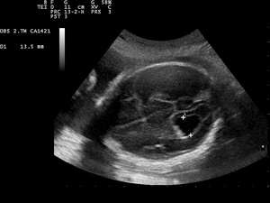
Ventriculomegaly
Ventriculomegaly is a brain condition that mainly occurs in the fetus when the lateral ventricles become dilated. The most common definition uses a width of the atrium of the lateral ventricle of greater than 10 mm.[1] This occurs in around 1% of pregnancies.[2] When this measurement is between 10 and 15 mm, the ventriculomegaly may be described as mild to moderate. When the measurement is greater than 15mm, the ventriculomegaly may be classified as more severe.[3] Enlargement of the ventricles may occur for a number of reasons, such as loss of brain volume (perhaps due to infection or infarction), or impaired outflow or absorption of cerebrospinal fluid from the ventricles, called hydrocephalus or normal pressure hydrocephalus associated with conspicuous brain sulcus. Often, however, there is no identifiable cause. The interventricular foramen may be congenitally malformed, or may have become obstructed by infection, hemorrhage, or rarely tumor, which may impair the drainage of cerebrospinal fluid, and thus accumulation in the ventricles. This diagnosis is generally found in routine fetal anomaly scans at 18–22 weeks gestation. It is one of the more common abnormal brain findings on prenatal ultrasound, occurring in around 1–2 per 1000 pregnancies.[4] In many cases of mild ventriculomegaly, however, there is resolution of ventriculomegaly during the pregnancy.
Associations
Ventriculomegaly is also known to be associated with other malformations such as agenesis of the corpus callosum, spina bifida, and heart defects. Fetuses with both isolated ventriculomegaly and with other anomalies have an increased risk of having a chromosomal abnormality, including that of Down syndrome.[3][5]
Many conditions associated with ventriculomegaly can be defined prior to birth, but the possibility remains of other anomalies (either structural, chromosomal or genetic) only being identified later in pregnancy or after birth.[6] Ventriculomegaly associated with abnormal findings and other structural malformations, often has an adverse prognosis, which ranges from disability (often mild) to death. However, in cases of mild isolated ventriculomegaly, there is around a 90% chance of a normal outcome.[5][7]
Increasingly, fetal magnetic resonance imaging is being considered as part of the assessment of pregnancies complicated by fetal ventriculomegaly,[8] and appears to be important in the postnatal assessment of affected children.[9]
Although evaluation of lateral ventricles dimensions is decisive for establishing a diagnosis of ventriculomegaly, the shape of the ventricular system, including that of the frontal horns, is also important.[10]
See also
References
- Cardoza JD, Goldstein RB, Filly RA. 1988. Exclusion of fetal ventriculomegaly with a single measurement: the width of the lateral ventricular atrium. Radiology 169: 711–714.
- Salomon, LH, Bernard JP, Ville Y. 2007. Reference ranges for fetal ventricular width: a non-normal approach . Ultrasound Obstet Gynecol (in press) Abstract
- Breeze AC, Alexander PM, Murdoch EM, Missfelder-Lobos HH, Hackett GA, Lees CC. 2007. Obstetric and neonatal outcomes in severe fetal ventriculomegaly. Prenat Diagn. 27(2):124-9 Abstract
- Achiron R, Schimmel M, Achiron A, Mashiach S. 1993. Fetal mild idiopathic lateral ventriculomegaly: is there a correlation with fetal trisomy? Ultrasound Obstet Gynecol 3: 89–92.
- Gaglioti P, Danelon D, Bontempo S, Mombro M, Cardaropoli S, Todros T. 2005. Fetal cerebral ventriculomegaly: outcome in 176 cases. Ultrasound Obstet Gynecol. Apr;25(4):372-7.
- Breeze AC, Dey PK, Lees CC, Hackett GA, Smith GCS, Murdoch EM. 2005. Obstetric and neonatal outcomes in apparently isolated mild fetal ventriculomegaly. J Perinat Med 33: 236–240 Abstract
- Signorelli M, Tiberti A, Valseriati D, Molin E, Cerri V, Groli C, Bianchi UA. 2004. Width of the fetal lateral ventricular atrium between 10 and 12 mm: a simple variation of the norm? Ultrasound Obstet Gynecol. Jan;23(1):14-8.
- Glenn OA, Barkovich AJ. 2006. Magnetic resonance imaging of the fetal brain and spine: an increasingly important tool in prenatal diagnosis, part 1. AJNR Am J Neuroradiol. Sep;27(8):1604-11.
- Falip C, Blanc N, Maes E, Zaccaria I, Oury JF, Sebag G, Garel C. 2007. Postnatal clinical and imaging follow-up of infants with prenatal isolated mild ventriculomegaly: a series of 101 cases. Pediatr Radiol. 2007 Oct;37(10):981-9. Abstract
- Glonek M; Kedzia A; Derkowski W Prenatal assessment of ventriculomegaly: an anatomical study. Medical science monitor : international medical journal of experimental and clinical research 2003;9(7):MT69-77.