Pericardial effusion
Pericardial effusion ("fluid around the heart") is an abnormal accumulation of fluid in the pericardial cavity. Because of the limited amount of space in the pericardial cavity, fluid accumulation leads to an increased intrapericardial pressure which can negatively affect heart function. A pericardial effusion with enough pressure to adversely affect heart function is called cardiac tamponade. Pericardial effusion usually results from a disturbed equilibrium between the production and re-absorption of pericardial fluid, or from a structural abnormality that allows fluid to enter the pericardial cavity.
| Pericardial effusion | |
|---|---|
.gif) | |
| A 2D transthoracic echocardiogram of pericardial effusion. The "swinging" heart | |
| Specialty | Cardiac surgery |
Normal levels of pericardial fluid are from 15 to 50 ml.
Signs and symptoms
Chest pain or pressure are common symptoms. A small effusion may be asymptomatic. Larger effusions may cause cardiac tamponade, a life-threatening complication; signs of impending tamponade include dyspnea, low blood pressure, and distant heart sounds.
The so-called "water-bottle heart" is a radiographic sign of pericardial effusion, in which the cardiopericardial silhouette is enlarged and assumes the shape of a flask or water bottle.
It can be associated with dullness to percussion over the left subscapular area due to compression of the left lung base. This phenomenon is known as Ewart's sign.[1]
Causes
- Pericarditis
- Long term usage of cabergoline ingredient (dopamine agonists)
- Viral infection (coxsackie virus)
- Infection including tuberculosis
- Drug eluting stents
- Inflammatory disorders, such as lupus, rheumatoid arthritis[2] and post myocardial infarction pericarditis (Dressler's syndrome)
- Cancer that has spread to the pericardium
- Trichinosis
- Kidney failure with excessive blood levels of urea nitrogen
- Minoxidil
- Hypothyroidism
- Heart surgery[3] (Postpericardiotomy syndrome)
Diagnosis
It may be:
- transudative (congestive heart failure, myxoedema, nephrotic syndrome),
- exudative (tuberculosis, spread from empyema)
- hemorrhagic (trauma, rupture of aneurysms, malignant effusion).
- malignant (due to fluid accumulation caused by metastasis)
The most common causes of pericardial effusion have changed over time and vary depending on geography and the population in question. When pericardial effusion is suspected, echocardiography usually confirms the diagnosis and allows assessment for signs of hemodynamic instability. Cross-sectional imaging with computed tomography (CT) can help to localize and quantify (as in a loculated effusion) or assess for pericardial pathology (pericardial thickening, constrictive pericarditis).[4]
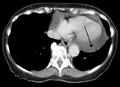 A CT scan image showing a pericardial effusion
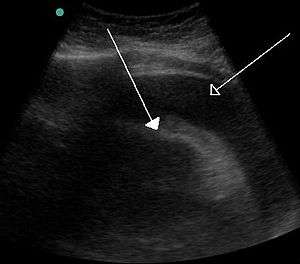 A very large hemorrhagic pericardial effusion due to malignancy as seen on ultrasound. closed arrow: the heart, open arrow: the effusion
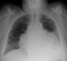
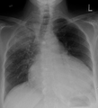 Pericardial effusion due to malignancy. Note bulbous heart and primary lung cancer in right upper lobe.
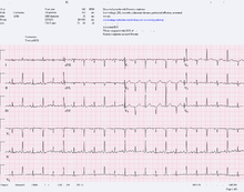
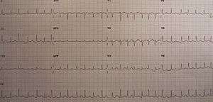 An ECG showing electrical alternans in a person with a pericardial effusion.
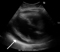 A pericardial effusion due to pericarditis
- Loculated pericardial effusion[5]
Treatment
Treatment depends on the underlying cause and the severity of the heart impairment. Pericardial effusion due to a viral infection usually goes away within a few weeks without the treatment. Some pericardial effusions remain small and never need treatment. If the pericardial effusion is due to a condition such as lupus, treatment with anti-inflammatory medications may help. If the effusion is compromising heart function and causing cardiac tamponade, it will need to be drained, most commonly by a needle inserted through the chest wall and into the pericardial space called pericardiocentesis. A drainage tube is often left in place for several days. In some cases, surgical drainage may be required by cutting through the pericardium creating a pericardial window.
References
- "Pericardial Disease".
- Hallewell RA, Sherratt DJ (1976). "Isolation and characterization of Co1E2 plasmid mutants unable to kill colicin-sensitive cells". Mol Gen Genet. 146 (3): 239–45. doi:10.1007/bf00701246. PMID 794689.
- Pericardial effusion:What are the symptoms?, Dr. Martha Grogan M.D.
- Chang, S (Jul–Sep 2014). "Brief Images: Massive pericardial effusion". Images in Paediatric Cardiology. 16 (3): 1–3. PMC 4521324. PMID 26236369.
- "UOTW #25 - Ultrasound of the Week". Ultrasound of the Week. 11 November 2014. Retrieved 27 May 2017.
External links
| Classification | |
|---|---|
| External resources |
- Pericardial Disease Cleveland Clinic Online Medical Reference