Fundus (eye)
The fundus of the eye is the interior surface of the eye opposite the lens and includes the retina, optic disc, macula, fovea, and posterior pole.[1] The fundus can be examined by ophthalmoscopy[1] and/or fundus photography.
| Fundus | |
|---|---|
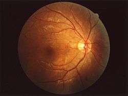 Fundus of human eye | |
| Identifiers | |
| MeSH | D005654 |
| Anatomical terminology | |
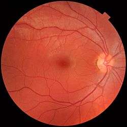
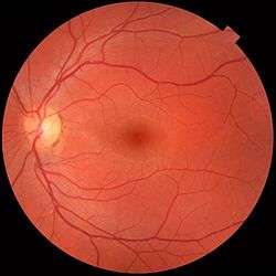
Each fundus has no sign of disease or pathology. The gaze is into the camera, so in each picture the macula is in the center of the image, and the optic disc is located towards the nose. Both optic discs have some pigmentation at the perimeter of the lateral side, which is considered non-pathological.
The left image (right eye) shows lighter areas close to larger vessels, which has been regarded as a normal finding in younger people.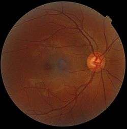
Variation
The color of the fundus varies both between and within species. In one study[2] of primates the retina is blue, green, yellow, orange, and red; only the human fundus (from a lightly pigmented blond person) is red. The major differences noted among the "higher" primate species were size and regularity of the border of macular area, size and shape of the optic disc, apparent 'texturing' of retina, and pigmentation of retina.
Clinical significance
Medical signs that can be detected from observation of eye fundus (generally by funduscopy) include hemorrhages, exudates, cotton wool spots, blood vessel abnormalities (tortuosity, pulsation and new vessels) and pigmentation.[3] Arteriolar constriction, seen as "silver wiring", and vascular tortuosities are seen in hypertensive retinopathy.
The eye's fundus is the only part of the human body where the microcirculation can be observed directly.[4] The diameter of the blood vessels around the optic disc is about 150 μm, and an ophthalmoscope allows observation of blood vessels with diameters as small as 10 μm.[4]
See also
| Wikimedia Commons has media related to Ocular fundus. |
References
- Cassin, B. and Solomon, S. Dictionary of Eye Terminology. Gainesville, Florida: Triad Publishing Company, 1990.
- Wolin LR, Massopust LC (September 1967). "Characteristics of the ocular fundus in primates". J. Anat. 101 (Pt 4): 693–9. PMC 1270903. PMID 6059819.Free full text in PubMed Central
- Imran Akram, Adrian Rubinstein "Common retinal signs. An overview", "Optometry Today", 28/01/05,
- Ronald Pitts Crick, Peng Tee Khaw, A Textbook of Clinical Ophthalmology: A Practical Guide to Disorders of the Eyes and Their Management, 3rd edition, World Scientific, 2003, ISBN 981-238-128-7
| Fibrous tunic (outer) |
|   | |||||
|---|---|---|---|---|---|---|---|
| Uvea/vascular tunic (middle) |
| ||||||
| Retina (inner) |
| ||||||
| Anatomical regions of the eye |
| ||||||
| Other | |||||||