Muscle spindle
Muscle spindles are stretch receptors within the body of a muscle that primarily detect changes in the length of the muscle. They convey length information to the central nervous system via afferent nerve fibers. This information can be processed by the brain as proprioception. The responses of muscle spindles to changes in length also play an important role in regulating the contraction of muscles, for example, by activating motor neurons via the stretch reflex to resist muscle stretch.
| Muscle spindle | |
|---|---|
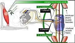 Mammalian muscle spindle showing typical position in a muscle (left), neuronal connections in spinal cord (middle) and expanded schematic (right). The spindle is a stretch receptor with its own motor supply consisting of several intrafusal muscle fibres. The sensory endings of a primary (group Ia) afferent and a secondary (group II) afferent coil around the non-contractile central portions of the intrafusal fibres. Gamma motoneurons activate the intrafusal muscle fibres, changing the resting firing rate and stretch-sensitivity of the afferents. [lower-alpha 1] | |
| Details | |
| Part of | Muscle |
| System | Musculoskeletal |
| Identifiers | |
| Latin | fusus neuromuscularis |
| MeSH | D009470 |
| TH | H3.11.06.0.00018 |
| FMA | 83607 |
| Anatomical terminology | |
The muscle spindle has both sensory and motor components.
- Sensory information conveyed by primary type Ia sensory fibers which spiral around muscle fibres within the spindle, and secondary type II sensory fibers
- Activation of muscle fibres within the spindle by up to a dozen gamma motor neurons and to a lesser extent by one or two beta motor neurons
Structure
Muscle spindles are found within the belly of muscles, between extrafusal muscle fibers.[lower-alpha 2] The specialised fibers that constitute the muscle spindle are known as intrafusal fibers (as they are present within the spindle), to distinguish themselves from the fibres of the muscle itself which are called extrafusal fibers. Muscle spindles have a capsule of connective tissue, and run parallel to the extrafusal muscle fibers.[lower-alpha 3]
Composition
Muscle spindles are composed of 5-14 muscle fibers, of which there are three types: dynamic nuclear bag fibers (bag1 fibers), static nuclear bag fibers (bag2 fibers), and nuclear chain fibers.[1][2]
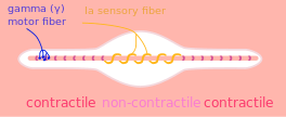
Primary type Ia sensory fibers (large diameter) spiral around all intrafusal muscle fibres, ending near the middle of each fibre. Secondary type II sensory fibers (medium diameter) end adjacent to the central regions of the static bag and chain fibres.[2] These fibres send information by stretch-sensitive mechanically-gated ion-channels of the axons.[3]
The motor part of the spindle is provided by motor neurons: up to a dozen gamma motor neurons and one or two beta motor neurons, collectively called fusimotor neurons. These activate the muscle fibres within the spindle. Gamma motor neurons supply only muscle fibres within the spindle, whereas beta motor neurons supply muscle fibres both within and outside of the spindle. Activation of the neurons causes a contraction and stiffening of the end parts of the muscle spindle muscle fibers.
Fusimotor neurons are classified as static or dynamic according to the type of muscle fibers they innervate and their effects on the responses of the Ia and II sensory neurons innervating the central, non-contractile part of the muscle spindle.
- The static axons innervate the chain or static bag2 fibers. They increase the firing rate of Ia and II afferents at a given muscle length (see schematic of fusimotor action below).
- The dynamic axons innervate the bag1 intrafusal muscle fibers. They increase the stretch-sensitivity of the Ia afferents by stiffening the bag1 intrafusal fibers.
Efferent nerve fibers of gamma motoneurons also terminate in muscle spindles; they make synapses at either or both of the ends of the intrafusal muscle fibers and regulate the sensitivity of the sensory afferents, which are located in the non-contractile central (equatorial) region.[4]
Function
Stretch reflex
When a muscle is stretched, primary type Ia sensory fibers of the muscle spindle respond to both changes in muscle length and velocity and transmit this activity to the spinal cord in the form of changes in the rate of action potentials. Likewise, secondary type II sensory fibers respond to muscle length changes (but with a smaller velocity-sensitive component) and transmit this signal to the spinal cord. The Ia afferent signals are transmitted monosynaptically to many alpha motor neurons of the receptor-bearing muscle. The reflexly evoked activity in the alpha motoneurons is then transmitted via their efferent axons to the extrafusal fibers of the muscle, which generate force and thereby resist the stretch. The Ia afferent signal is also transmitted polysynaptically through interneurons (Ia inhibitory interneurons), which inhibit alpha motorneurons of antagonist muscles, causing them to relax.
Sensitivity modification
The function of the gamma motor neurons is not to supplement the force of muscle contraction provided by the extrafusal fibers, but to modify the sensitivity of the muscle spindle sensory afferents to stretch. Upon release of acetylcholine by the active gamma motor neuron, the end portions of the intrafusal muscle fibers contract, thus elongating the non-contractile central portions (see "fusimotor action" schematic below). This opens stretch-sensitive ion channels of the sensory endings, leading to an influx of sodium ions. This raises the resting potential of the endings, thereby increasing the probability of action potential firing, thus increasing the stretch-sensitivity of the muscle spindle afferents.
How does the central nervous system control gamma fusimotor neurons? It has been difficult to record from gamma motoneurons during normal movement because they have very small axons. Several theories have been proposed, based on recordings from spindle afferents.
- 1) Alpha-gamma coactivation. Here it is posited that gamma motoneurons are activated in parallel with alpha motoneurons to maintain the firing of spindle afferents when the extrafusal muscles shorten.[5]
- 2) Fusimotor set: Gamma motoneurons are activated according to the novelty or difficulty of a task. Whereas static gamma motoneurons are continuously active during routine movements such as locomotion, dynamic gamma motoneoruns tend to be activated more during difficult tasks, increasing Ia stretch-sensitivity.[6]
- 3) Fusimotor template of intended movement. Static gamma activity is a "temporal template" of the expected shortening and lengthening of the receptor-bearing muscle. Dynamic gamma activity turns on and off abruptly, sensitizing spindle afferents to the onset of muscle lengthening and departures from the intended movement trajectory.[7]
Development
It is also believed that muscle spindles play a critical role in sensorimotor development.
Clinical significance
After stroke or spinal cord injury in humans, spastic hypertonia (spastic paralysis) often develops, whereby the stretch reflex in flexor muscles of the arms and extensor muscles of the legs is overly sensitive. This results in abnormal postures, stiffness and contractures. Hypertonia may be the result of over-sensitivity of alpha motoneurons and interneurons to the Ia and II afferent signals.[8]
Additional images
 Muscle spindle
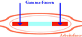 Gamma fiber
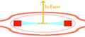 1A fiber
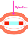 Alpha fiber
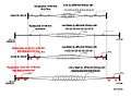 schematic of fusimotor action
See also
Notes
- Animated version: https://www.ualberta.ca/~aprochaz/research_interactive_receptor_model.html Arthur Prochazka's Lab, University of Alberta
- "Fusus" Latin: "spindle"
- unlike Golgi tendon organs, which are oriented in series
References
- Mancall, Elliott L; Brock, David G, eds. (2011). "Chapter 2 - Overview of the Microstructure of the Nervous System". Gray’s Clinical Neuroanatomy: The Anatomic Basis for Clinical Neuroscience. Elsevier Saunders. pp. 29–30. ISBN 978-1-4160-4705-6.
- Pearson, Keir G; Gordon, James E (2013). "35 - Spinal Reflexes". In Kandel, Eric R; Schwartz, James H; Jessell, Thomas M; Siegelbaum, Steven A; Hudspeth, AJ (eds.). Principles of Neural Science (5th ed.). United State of America: McGraw-Hill. pp. 794–795. ISBN 978-0-07-139011-8.
- Purves, Dale; Augustine, George J; Fitzpatrick, David; Hall, William C; Lamantia, Anthony Samuel; Mooney, Richard D; Platt, Michael L; White, Leonard E, eds. (2018). "Chapter 9 - The Somatosensory System: Touch and Proprioception". Neuroscience (6th ed.). Sinauer Associates. pp. 201–202. ISBN 9781605353807.
- Hulliger M (1984). The mammalian muscle spindle and its central control. Rev. Physiol. Biochem. Pharmacol. Reviews of Physiology, Biochemistry and Pharmacology. 101. pp. 1–110. doi:10.1007/bfb0027694. ISBN 978-3-540-13679-8. PMID 6240757.
- Vallbo AB, al-Falahe NA (February 1990). "Human muscle spindle response in a motor learning task". J. Physiol. 421: 553–68. doi:10.1113/jphysiol.1990.sp017961. PMC 1190101. PMID 2140862.
- Prochazka, A. (1996). "Proprioceptive feedback and movement regulation". In Rowell, L.; Sheperd, J.T. (eds.). Exercise: Regulation and Integration of Multiple Systems. Handbook of physiology. New York: American Physiological Society. pp. 89–127. ISBN 978-0195091748.
- Taylor A, Durbaba R, Ellaway PH, Rawlinson S (March 2006). "Static and dynamic gamma-motor output to ankle flexor muscles during locomotion in the decerebrate cat". J. Physiol. 571 (Pt 3): 711–23. doi:10.1113/jphysiol.2005.101634. PMC 1805796. PMID 16423858.
- Heckmann CJ, Gorassini MA, Bennett DJ (February 2005). "Persistent inward currents in motoneuron dendrites: implications for motor output". Muscle Nerve. 31 (2): 135–56. CiteSeerX 10.1.1.126.3583. doi:10.1002/mus.20261. PMID 15736297.
External links
- Muscle+Spindles at the US National Library of Medicine Medical Subject Headings (MeSH)
| Authority control |
|---|