Muscle tissue
Muscle tissue is a soft tissue that composes muscles in animal bodies, and gives rise to muscles' ability to contract. This is opposed to other components or tissues in muscle such as tendons or perimysium. It is formed during embryonic development through a process known as myogenesis.[1] Muscle tissue consists of elongated cells also called as muscle fibres. This tissue is responsible for movements in our body. Muscles contain special proteins called contractile protein which contract and relax to cause movement
| Muscle tissue | |
|---|---|
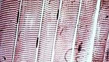
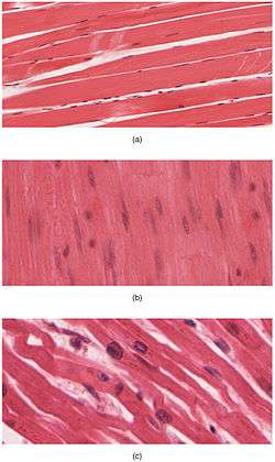 The body contains three types of muscle tissue: (a) skeletal muscle, (b) smooth muscle, and (c) cardiac muscle. (Same magnification) | |
.svg.png) A schematic diagram of the different types of muscle cells (same order as above). | |
| Anatomical terminology |
Muscle tissues varies with function and location in the body. In mammals the three types are: skeletal or striated muscle; smooth or non-striated muscle; and cardiac muscle, which is sometimes known as semi-striated. Smooth and cardiac muscle contracts involuntarily, without conscious intervention. These muscle types may be activated both through interaction of the central nervous system as well as by receiving innervation from peripheral plexus or endocrine (hormonal) activation. Striated or skeletal muscle only contracts voluntarily, upon influence of the central nervous system. Reflexes are a form of nonconscious activation of skeletal muscles, but nonetheless arise through activation of the central nervous system, albeit not engaging cortical structures until after the contraction has occurred.[1]
The different muscle types vary in their response to neurotransmitters and endocrine substances such as acetylcholine, noradrenaline, adrenaline, nitric oxide and among others depending on muscle type and the exact location of the muscle.[1]
Sub-categorization of muscle tissue is also possible, depending on among other things the content of myoglobin, mitochondria, myosin ATPase etc.
Structure
Muscle tissue :D is an elongated tissue ranging from several millimetres to about 10 centimetres in length and from 10 to 100 micrometres in width.tispotter/muscle_tissue.htm |date=2014-10-21 }}</ref> These cells are joined together in tissues that may be either striated or smooth, depending on the presence or absence, respectively, of organized, regularly repeated arrangements of myofibrillar contractile proteins called myofilaments. Striated muscle is further classified as either skeletal or cardiac muscle.[2] Striated muscle is typically subject to conscious control, while smooth muscle is not. Thus, muscle tissue can be described as being one of three different types:
- Skeletal muscle, striated in structure and under voluntary control, is anchored by tendons (or by aponeuroses at a few places) to bone and is used to effect skeletal movement such as locomotion and to maintain posture. (Though postural control is generally maintained as an unconscious reflex—see proprioception—the muscles responsible also react to conscious control like non-postural muscles.) An average adult male is made up of 42% of skeletal muscle and an average adult female is made up of 36% (as a percentage of body mass).[3] It also has striations unlike smooth muscle.
- Smooth muscle, neither striated in structure nor under voluntary control, is found within the walls of organs and structures such as the esophagus, stomach, intestines, bronchi, uterus, urethra, bladder, blood vessels, and the arrector pili in the skin (in which it controls erection of body hair).
In vertebrates, there is a third muscle tissue recognized:
- Cardiac muscle (myocardium), found only in the heart, is a striated muscle similar in structure to skeletal muscle but not subject to voluntary control.
Cardiac and skeletal muscles are "striated" in that they contain sarcomeres and are packed into highly regular arrangements of bundles; smooth muscle has neither. While skeletal muscles are arranged in regular, parallel bundles, cardiac muscle connects at branching, irregular angles (called intercalated discs). Striated muscle contracts and relaxes in short, intense bursts, whereas smooth muscle sustains longer or even near-permanent contractions.
Comparison of types
| smooth muscle | cardiac muscle | skeletal muscle | |
| Anatomy | |||
| Neuromuscular junction | none | none | present |
| Fibers | fusiform, short (<0.4 mm) | branching | cylindrical, long (<15 cm) |
| Mitochondria | few | numerous | many to few (by type) |
| Nuclei | 1 | 1 | >1 |
| Sarcomeres | none | present, max. length 2.6 µm | present, max. length 3.7 µm |
| Syncytium | none (independent cells) | none (but functional as such) | present |
| Sarcoplasmic reticulum | little elaborated | moderately elaborated | highly elaborated |
| ATPase | little | moderate | abundant |
| Physiology | |||
| Self-regulation | spontaneous action (slow) | yes (rapid) | none (requires nerve stimulus) |
| Response to stimulus | unresponsive | "all-or-nothing" | "all-or-nothing" |
| Action potential | yes | yes | yes |
| Workspace | Force/length curve is variable | the increase in the force/length curve | at the peak of the force/length curve |
| Response to stimulus | 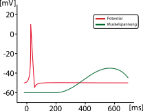 |
 |
 |
Skeletal muscle
Skeletal muscle is further divided into several subtypes:
- Type I, slow oxidative, slow twitch, or "red" muscle is dense with capillaries and is rich in mitochondria and myoglobin, giving the muscle tissue its characteristic red color. It can carry more oxygen and sustain aerobic activity.
- Type I muscle fiber are sometimes broken down into Type I and Type Ic categories, as a result of recent research.
- Type II, fast twitch muscle, has three major kinds that are, in order of increasing contractile speed:[4]
- Type IIa, which, like slow muscle, is aerobic, rich in mitochondria and capillaries and appears red when deoxygenated.
- Type IIx (also known as type IId), which is less dense in mitochondria and myoglobin. This is the fastest muscle type in humans. It can contract more quickly and with a greater amount of force than oxidative muscle, but can sustain only short, anaerobic bursts of activity before muscle contraction becomes painful (often incorrectly attributed to a build-up of lactic acid). N.B. in some books and articles this muscle in humans was, confusingly, called type IIB.[5]
- Type IIb, which is anaerobic, glycolytic, "white" muscle that is even less dense in mitochondria and myoglobin. In small animals like rodents this is the major fast muscle type, explaining the pale color of their flesh.
Smooth muscle
Smooth muscle is an involuntary non-striated muscle. It is divided into two subgroups: the single-unit (unitary) and multiunit smooth muscle. Within single-unit cells, the whole bundle or sheet contracts as a syncytium (i.e. a multinucleate mass of cytoplasm that is not separated into cells). Multiunit smooth muscle tissues innervate individual cells; as such, they allow for fine control and gradual responses, much like motor unit recruitment in skeletal muscle.
Smooth muscle is found within the walls of blood vessels (such smooth muscle specifically being termed vascular smooth muscle) such as in the tunica media layer of large (aorta) and small arteries, arterioles and veins. Smooth muscle is also found in lymphatic vessels, the urinary bladder, uterus (termed uterine smooth muscle), male and female reproductive tracts, gastrointestinal tract, respiratory tract, arrector pili of skin, the ciliary muscle, and iris of the eye. The structure and function is basically the same in smooth muscle cells in different organs, but the inducing stimuli differ substantially, in order to perform individual effects in the body at individual times. In addition, the glomeruli of the kidneys contain smooth muscle-like cells called mesangial cells.
Cardiac muscle
Cardiac muscle is involuntary, striated muscle that is found in the walls and histological foundation of the heart, specifically the myocardium. Cardiac muscle is one of three major types of muscle, the others being skeletal and smooth muscle. These three types of muscle all form in the process of myogenesis. The cells that constitute cardiac muscle, called cardiomyocytes or myocardiocytes, predominantly contain only one nucleus, although populations with two to four nuclei do exist.[6][7] The myocardium is the muscle tissue of the heart, and forms a thick middle layer between the outer epicardium layer and the inner endocardium layer.
Coordinated contractions of cardiac muscle cells in the heart propel blood out of the atria and ventricles to the blood vessels of the left/body/systemic and right/lungs/pulmonary circulatory systems. This complex mechanism illustrates systole of the heart.
Cardiac muscle cells, unlike most other tissues in the body, rely on an available blood and electrical supply to deliver oxygen and nutrients and remove waste products such as carbon dioxide. The coronary arteries help fulfill this function.
Function
Smooth muscle
Smooth musculature is found in almost all organ systems such as hollow organs (e.g. stomach, bladder), in tubular structures (e.g. vessels, bile ducts), in sphincters, in the uterus, in the eye etc. In addition it plays an important role in the ducts of exocrine glands. It fulfills various tasks such as sealing orifices (e.g. pylorus, uterine os) or the transport of the chyme through wavelike contractions of the intestinal tube. On the one hand smooth muscle cells contract slower than skeletal muscle cells, on the other hand they are stronger, more sustained and require less energy.
Cardiac muscle
Cardiac muscle is the muscle of the heart. It is self-contracting, autonomically regulated and must continue to contract in rhythmic fashion for the whole life of the organism. Hence it has special features.
See also
References
- "Muscle Tissue TheVisualMD.com". www.thevisualmd.com. Retrieved 2015-12-30.
- Pratt, Rebecca. "Muscle Tissue". AnatomyOne. Amirsys, Inc. Archived from the original on 2 February 2017. Retrieved 26 October 2012.
- Marieb, Elaine; Hoehn, Katja (2007). Human Anatomy & Physiology (7th ed.). Pearson Benjamin Cummings. p. 317. ISBN 978-0-8053-5387-7.
- Larsson, L; Edström, L; Lindegren, B; Gorza, L; Schiaffino, S (July 1991). "MHC composition and enzyme-histochemical and physiological properties of a novel fast-twitch motor unit type". The American Journal of Physiology. 261 (1 pt 1): C93–101. doi:10.1152/ajpcell.1991.261.1.C93. PMID 1858863.
- Smerdu, V; Karsch-Mizrachi, I; Campione, M; Leinwand, L; Schiaffino, S (December 1994). "Type IIx myosin heavy chain transcripts are expressed in type IIb fibers of human skeletal muscle". The American Journal of Physiology. 267 (6 pt 1): C1723–1728. doi:10.1152/ajpcell.1994.267.6.C1723. PMID 7545970. Note: Access to full text requires subscription; abstract freely available
- Olivetti G, Cigola E, Maestri R, et al. (July 1996). "Aging, cardiac hypertrophy and ischemic cardiomyopathy do not affect the proportion of mononucleated and multinucleated myocytes in the human heart". Journal of Molecular and Cellular Cardiology. 28 (7): 1463–77. doi:10.1006/jmcc.1996.0137. PMID 8841934.
- Pollard, Thomas D. and Earnshaw, William. C., "Cell Biology". Philadelphia: Saunders. 2007.
| Animals | |
|---|---|
| Plants |
|
| |