Intrafusal muscle fiber
Intrafusal muscle fibers are skeletal muscle fibers that serve as specialized sensory organs (proprioceptors) that detect the amount and rate of change in length of a muscle.[1] They constitute the muscle spindle and are innervated by both sensory (afferent) and motor (efferent) fibers. Gamma efferents from small multipolar cells from anterior gray column innervate it. These form a part of neuromuscular spindles. Intrafusal muscle fibers are walled off from the rest of the muscle by an outer connective tissue sheath consisting of flattened fibroblasts and collagen.[2] This sheath has a spindle or "fusiform" shape, hence the name "intrafusal".
| Intrafusal muscle fiber | |
|---|---|
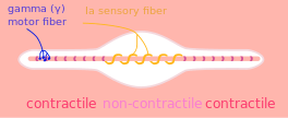 A muscle spindle, with γ motor and Ia sensory fibers | |
| Details | |
| Part of | Skeletal muscle |
| Function | Proprioception |
| Identifiers | |
| Latin | myofibra intrafusalis |
| TH | H3.03.00.0.00012 |
| Anatomical terms of microanatomy | |
There are two types of intrafusal muscle fibers: nuclear bag and nuclear chain fibers. They bear two types of sensory ending, known as annulospiral and flower-spray endings. Both ends of these fibers contract but the central region only stretches and does not contract.
They are innervated by gamma motor neurons and beta motor neurons.
It is by the sensory information from these two intrafusal fiber types that an individual is able to judge the position of their muscle, and the rate at which it is changing.
Intrafusal muscle fibers are not to be confused with extrafusal muscle fibers, which contract, generating skeletal movement and are innervated by alpha motor neurons.
See also
References
- Casagrand, Janet (2008) Action and Movement: Spinal Control of Motor Units and Spinal Reflexes. University of Colorado, Boulder.
- Mancall, Elliott L; Brock, David G, eds. (2011). "Chapter 2 - Overview of the Microstructure of the Nervous System". Gray’s Clinical Neuroanatomy: The Anatomic Basis for Clinical Neuroscience. Elsevier Saunders. pp. 29–30. ISBN 978-1-4160-4705-6.
External links
- Histology at ucsd.edu
- Histology at umdnj.edu
- "Chapter 1: The Muscle Spindle and the Central Nervous System". Neuromuscular Reeducation with Electromyometric Feedback (PDF). Advanced Therapy Institute. Retrieved 30 November 2013.
| Authority control |
|---|