Habenula
In neuroanatomy, habenula (diminutive of Latin habena meaning rein) originally denoted the stalk of the pineal gland (pineal habenula; pedunculus of pineal body), but gradually came to refer to a neighboring group of nerve cells with which the pineal gland was believed to be associated, the habenular nucleus. The habenular nucleus is a set of well-conserved structures in all vertebrate animals.[1]
| Habenula | |
|---|---|
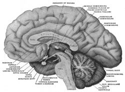 Mesal aspect of a brain sectioned in the median sagittal plane. Habenula is not labeled directly, but after expanding, look to region with 'habenular commissure', 'pineal body', and 'posterior commissure' | |
| |
| Identifiers | |
| MeSH | D019262 |
| NeuroNames | 294 |
| NeuroLex ID | birnlex_1611 |
| TA | A14.1.08.003 |
| FMA | 62032 |
| Anatomical terms of neuroanatomy | |
Currently, the Terminologia Anatomica term refers exclusively to this separate cell mass in the caudal and dorsal aspect of the dorsal thalamus (the epithalamus), embedded in the posterior end of the medullary stria from which it receives most of its afferent fibers. By way of the fasciculus retroflexus (habenulointerpeduncular tract) it projects to the interpeduncular nucleus and other paramedian cell groups of the midbrain tegmentum.
The habenula receives input via the stria medullaris thalami and outputs to many midbrain areas involved in releasing neurotransmitters, such as dopamine, norepinephrine, and serotonin.
Anatomy
The habenula was traditionally divided into lateral (limbic) and medial (motor) parts. Detailed examination of the region in the cat, however, suggested that the lateral part should be further divided into ten distinct subnuclei and the medial into five distinct subnuclei.[2]
Asymmetry
Various species exhibit left-right asymmetric differentiation of habenular neurons. In many fishes and amphibians, the habenula on one side is significantly larger and better organized into distinct nuclei in the dorsal diencephalon than its smaller pair. The sidedness of such differentiation (whether the left or the right is more developed) varies with the species. In humans, however, both habenulae are symmetrically small and poorly developed.[3]
Lateral habenula
The primary input regions to the lateral habenula (LHb) are the lateral preoptic area (bringing input from the hippocampus and lateral septum), the ventral pallidum (bringing input from the nucleus accumbens and mediodorsal nucleus of the thalamus), the lateral hypothalamus, the medial habenula, and the internal segment of the globus pallidus (bringing input from other basal ganglia structures).[4]
Neurons in the lateral habenula are ‘reward-negative’ as they are activated by stimuli associated with unpleasant events, the absence of the reward or the presence of punishment especially when this is unpredictable.[5] Reward information to the lateral habenula comes from the internal part of the globus pallidus.[6]
The outputs of the lateral habenula target dopaminergic regions (substantia nigra pars compacta and the ventral tegmental area), serotonergic regions (median raphe and dorsal raphe nuclei), and a cholinergic region (the laterodorsal tegmental nucleus).[4] This output inhibits dopamine neurons in substantia nigra pars compacta and the ventral tegmental area, with activation in the lateral habenula linking to deactivation in them, and vice versa, deactivation in the lateral habenula with their activation.[7] The lateral habenula functions to oppose the action of the laterodorsal tegmental nucleus in the acquisition of avoidance responses but not the processing of avoidance later on when it is a memory, motivation or its execution.[8] New research suggests that lateral habenula may play a crucial role in decision making.[9]
Medial habenula
Input to the medial habenula (MHb) comes from a variety of regions and carries a number of different chemicals. Input regions include septal nuclei (the nucleus fimbrialis septi and the nucleus triangularis septi), dopaminergic inputs from the interfascicular nucleus of the ventral tegmental area, noradrenergic inputs from the locus ceruleus, and GABAergic inputs from the diagonal band of Broca. The medial habenula sends outputs of glutamate, substance P and acetylcholine to the periaqueductal gray via the interpeduncular nucleus as well as to the pineal gland.[10][11]
Olfactory coding in the habenula
In lower vertebrates (lampreys and teleost fishes), mitral cell (principal olfactory neurons) axons project exclusively to the right hemisphere of the habenula in an asymmetric manner. It is reported that the dorsal habenulae (DHb) are functionally asymmetric with predominantly odor responses in the right hemisphere. It was also shown that DHb neurons are spontaneously active even in the absence of olfactory stimulation. These spontaneously-active DHb neurons are organized into functional clusters which were proposed to govern olfactory responses. (Jetti, SK. et al 2014, Current Biology)
Functions
The habenular nuclei are involved in pain processing, reproductive behavior, nutrition, sleep-wake cycles, stress responses, and learning.[1] Recent demonstrations using fMRI[12] and single unit electrophysiology[7] have closely linked the function of the lateral habenula with reward processing, in particular with regard to encoding negative feedback or negative rewards. Matsumoto and Hikosaka suggested in 2007 that this reward and reward-negative information in the brain might "be elaborated through the interplay among the lateral habenula, the basal ganglia, and monoaminergic (dopaminergic and serotonergic) systems" and that the lateral habenula may play a pivotal role in this "integrative function".[7] Recent evidence suggests that neurons in the lateral habenula signal positive and negative information-prediction errors in addition to positive and negative reward-prediction errors.[13]
Depression
Both the medial and lateral habenula show reduced volume in those with depression. Neuron cell numbers were also reduced on the right side.[14] Such changes are not seen in those with schizophrenia.[14] Deep brain stimulation of the major afferent bundle (i.e., stria medullaris thalami) of the lateral habenula has been used for treatment of depression where it is severe, protracted and therapy-resistant.[15][16]
Methyl-D-aspartate (NMDA) receptor-dependent burst firing in the lateral habenula has been associated with depression in animal studies,[17] and it has been shown that the general anesthetic ketamine blocks this firing by acting as a receptor antagonist.[18] Ketamine has been the subject of numerous studies after having shown fast-acting antidepressant effects in humans.[19]
References
- Andres KH, von Düring M, Veh RW (April 1999). "Subnuclear organization of the rat habenular complexes". The Journal of Comparative Neurology. 407 (1): 130–50. doi:10.1002/(SICI)1096-9861(19990428)407:1<130::AID-CNE10>3.0.CO;2-8. PMID 10213193.
- Iwahori N (February 1977). "A Golgi study on the habenular nucleus of the cat". The Journal of Comparative Neurology. 72 (3): 319–44. doi:10.1002/cne.901710303. hdl:2433/221270. PMID 319124.
- Hüsken U, Stickney HL, Gestri G, Bianco IH, Faro A, Young RM, Roussigne M, Hawkins TA, Beretta CA, Brinkmann I, Paolini A, Jacinto R, Albadri S, Dreosti E, Tsalavouta M, Schwarz Q, Cavodeassi F, Barth AK, Wen L, Zhang B, Blader P, Yaksi E, Poggi L, Zigman M, Lin S, Wilson SW, Carl M (October 2014). "Tcf7l2 is required for left-right asymmetric differentiation of habenular neurons". Current Biology. 24 (19): 2217–27. doi:10.1016/j.cub.2014.08.006. PMC 4194317. PMID 25201686.
- Geisler S, Trimble M (June 2008). "The lateral habenula: no longer neglected". CNS Spectrums. 13 (6): 484–9. doi:10.1017/S1092852900016710. PMID 18567972.
- Matsumoto M, Hikosaka O (January 2009). "Representation of negative motivational value in the primate lateral habenula". Nature Neuroscience. 12 (1): 77–84. doi:10.1038/nn.2233. PMC 2737828. PMID 19043410.
- Hong S, Hikosaka O (November 2008). "The globus pallidus sends reward-related signals to the lateral habenula". Neuron. 60 (4): 720–9. doi:10.1016/j.neuron.2008.09.035. PMC 2638585. PMID 19038227.
- Matsumoto M, Hikosaka O (June 2007). "Lateral habenula as a source of negative reward signals in dopamine neurons". Nature. 447 (7148): 1111–5. Bibcode:2007Natur.447.1111M. doi:10.1038/nature05860. PMID 17522629.
- Shumake J, Ilango A, Scheich H, Wetzel W, Ohl FW (April 2010). "Differential neuromodulation of acquisition and retrieval of avoidance learning by the lateral habenula and ventral tegmental area". The Journal of Neuroscience. 30 (17): 5876–83. doi:10.1523/JNEUROSCI.3604-09.2010. PMC 6632612. PMID 20427648.
- Stopper CM, Floresco SB (January 2014). "What's better for me? Fundamental role for lateral habenula in promoting subjective decision biases". Nature Neuroscience. 17 (1): 33–5. doi:10.1038/nn.3587. PMC 4974073. PMID 24270185. Lay summary – Science Daily.
- Lecourtier L, Kelly PH (January 2007). "A conductor hidden in the orchestra? Role of the habenular complex in monoamine transmission and cognition". Neuroscience and Biobehavioral Reviews. 31 (5): 658–72. doi:10.1016/j.neubiorev.2007.01.004. PMID 17379307.
- Antolin-Fontes B, Ables JL, Görlich A, Ibañez-Tallon I (September 2015). "The habenulo-interpeduncular pathway in nicotine aversion and withdrawal". Neuropharmacology. 96 (Pt B): 213–22. doi:10.1016/j.neuropharm.2014.11.019. PMC 4452453. PMID 25476971.
- Ullsperger M, von Cramon DY (May 2003). "Error monitoring using external feedback: specific roles of the habenular complex, the reward system, and the cingulate motor area revealed by functional magnetic resonance imaging". The Journal of Neuroscience. 23 (10): 4308–14. doi:10.1523/JNEUROSCI.23-10-04308.2003. PMC 6741115. PMID 12764119.
- Bromberg-Martin ES, Hikosaka O (August 2011). "Lateral habenula neurons signal errors in the prediction of reward information". Nature Neuroscience. 14 (9): 1209–16. doi:10.1038/nn.2902. PMC 3164948. PMID 21857659.
- Ranft K, Dobrowolny H, Krell D, Bielau H, Bogerts B, Bernstein HG (April 2010). "Evidence for structural abnormalities of the human habenular complex in affective disorders but not in schizophrenia". Psychological Medicine. 40 (4): 557–67. doi:10.1017/S0033291709990821. PMID 19671211.
- Sartorius A, Kiening KL, Kirsch P, von Gall CC, Haberkorn U, Unterberg AW, Henn FA, Meyer-Lindenberg A (January 2010). "Remission of major depression under deep brain stimulation of the lateral habenula in a therapy-refractory patient". Biological Psychiatry. 67 (2): e9–e11. doi:10.1016/j.biopsych.2009.08.027. PMID 19846068.
- Juckel G, Uhl I, Padberg F, Brüne M, Winter C (February 2009). "Psychosurgery and deep brain stimulation as ultima ratio treatment for refractory depression". European Archives of Psychiatry and Clinical Neuroscience. 259 (1): 1–7. doi:10.1007/s00406-008-0826-7. PMID 19137233.
- Howe WM, Kenny PJ (February 2018). "Burst firing sets the stage for depression". Nature. 554 (7692): 304–305. doi:10.1038/d41586-018-01588-z. PMID 29446408.
- Yang Y, Cui Y, Sang K, Dong Y, Ni Z, Ma S, Hu H (February 2018). "Ketamine blocks bursting in the lateral habenula to rapidly relieve depression". Nature. 554 (7692): 317–322. Bibcode:2018Natur.554..317Y. doi:10.1038/nature25509. PMID 29446381.
- Serafini G, Howland RH, Rovedi F, Girardi P, Amore M (September 2014). "The role of ketamine in treatment-resistant depression: a systematic review". Current Neuropharmacology. 12 (5): 444–61. doi:10.2174/1570159X12666140619204251. PMC 4243034. PMID 25426012.
External links
- Stained brain slice images which include the "Habenula" at the BrainMaps project
- NIF Search - Habenula via the Neuroscience Information Framework
- Jetti SK, Vendrell-Llopis N, Yaksi E (February 2014). "Spontaneous activity governs olfactory representations in spatially organized habenular microcircuits". Current Biology. 24 (4): 434–9. doi:10.1016/j.cub.2014.01.015. PMID 24508164.
| Authority control |
|---|