Interpeduncular fossa
The interpeduncular fossa is a somewhat rhomboid-shaped area of the base of the brain, limited in front by the optic chiasma, behind by the antero-superior surface of the pons, antero-laterally by the converging optic tracts, and postero-laterally by the diverging cerebral peduncles.[1]
| Interpeduncular fossa | |
|---|---|
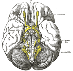 Base of brain | |
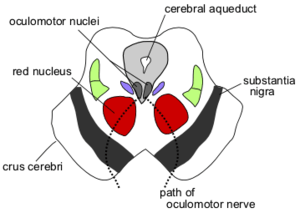 Section through superior colliculus showing path of oculomotor nerve (interpeduncular fossa not labeled, but visible at bottom center) | |
| Details | |
| Identifiers | |
| Latin | fossa interpeduncularis |
| NeuroNames | 489 |
| Anatomical terms of neuroanatomy | |
The floor of interpeduncular fossa, from behind forward, are the posterior perforated substance, corpora mamillaria, tuber cinereum, infundibulum, and Pituitary Gland.
Contents of interpeduncular fossa include oculomotor nerve, and circle of willis.
Anatomy
The interpeduncular fossa is located in the posterior portion of the brain, in the brain stem.
It has been found in humans and macaques, but not in rats or mice, showing that this is a relatively new evolutionary region.[2]
Clinical Significance
The most common locations for neurocutaneous melanosis have occurred along the interpeduncular fossa, ventral brainstem, upper cervical cord, and ventral lumbosacral cord.[3]
See also
Additional images
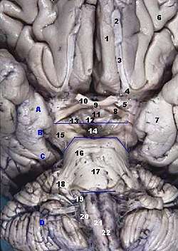 Human brainstem anterior view
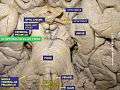 Interpeduncular fossa
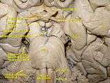 Cerebrum. Deep dissection. Inferior dissection.
References
This article incorporates text in the public domain from page 816 of the 20th edition of Gray's Anatomy (1918)
- "Interpeduncular fossa". IMAIOS. Retrieved 2019-04-03.
- "BrainInfo". braininfo.rprc.washington.edu. Retrieved 2019-04-03.
- Islam, Monica P. (2015). "Neurocutaneous melanosis". Neurocutaneous Syndromes. Handbook of Clinical Neurology. 132. pp. 111–7. doi:10.1016/B978-0-444-62702-5.00007-X. ISBN 978-0-444-62702-5. PMID 26564074.