Medial longitudinal fasciculus
The medial longitudinal fasciculus (MLF) is one of a pair of crossed over tracts, on each side of the brainstem. These bundles of axons are situated near the midline of the brainstem and are made up of both ascending and descending fibers that arise from a number of sources and terminate in different areas. The MLF is the main central connection for the oculomotor nerve, trochlear nerve, and abducens nerve. The vertical gaze center is at the rostral interstitial nucleus (riMLF).
| Medial longitudinal fasciculus | |
|---|---|
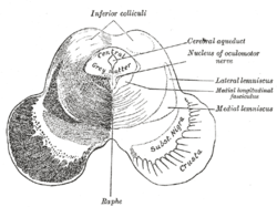 Transverse section of mid-brain at level of inferior colliculi. (Medial longitudinal fasciculus labeled at center right.) | |
 Axial section through mid-brain. 1. Corpora quadrigemina. 2. Cerebral aqueduct. 3. Central gray stratum. 4. Interpeduncular space. 5. Sulcus lateralis. 6. Substantia nigra. 7. Red nucleus of tegmentum. 8. Oculomotor nerve, with 8’, its nucleus oforigin. a. Lemniscus (in blue) with a’ the medial lemniscus and a" the lateral lemniscus. b. Medial longitudinal fasciculus. c. Raphe. d. Temporopontine fibers. e. Portion of medial lemniscus, which runs to the lentiform nucleus and insula. f. Cerebrospinal fibers. g. Frontopontine fibers. | |
| Details | |
| Identifiers | |
| Latin | fasciculus longitudinalis medialis |
| NeuroNames | 1588, 784 |
| NeuroLex ID | nlx_144065 |
| TA | A14.1.04.113 A14.1.05.304 A14.1.06.209 |
| FMA | 83846 |
| Anatomical terms of neuroanatomy | |
The MLF ascends to the interstitial nucleus of Cajal, which lies in the lateral wall of the third ventricle, just above the cerebral aqueduct.
Function
The medial longitudinal fasciculus carries information about the direction that the eyes should move.
It connects the cranial nerve nuclei III (Oculomotor nerve), IV (Trochlear nerve) and VI (Abducens nerve) together, and integrates movements directed by the gaze centers (frontal eye field) and information about head movement (from cranial nerve VIII, Vestibulocochlear nerve). It is an integral component of saccadic eye movements as well as vestibulo-ocular and optokinetic reflexes.
It also carries the descending tectospinal tract and medial vestibulospinal tracts into the cervical spinal cord, and innervates some muscles of the neck and upper limbs.
Clinical significance
A lesion of the MLF produces slowed or absent adduction of the ipsilateral eye, usually associated with involuntary jerky eye movements (nystagmus) of the abducting eye, a syndrome called internuclear ophthalmoplegia. Because multiple sclerosis causes demyelination of the axons of CNS, it can cause internuclear ophthalmoplegia when MLF axons get demyelinated,[1] where it presents as pathologic nystagmus and diplopia.
Inputs
The ascending MLF mainly arises from the superior and medial vestibular nucleus (VN) and is involved in the generation of the vestibulo-ocular reflex (VOR). This is achieved by inputs to the VN from:
- the vestibulocochlear (8th cranial) nerve about head movements,
- gait adjustments from the flocculus of the cerebellum,
- head and neck proprioceptors and foot and ankle muscle spindle, via the fastigial nucleus.
Descending fibers can also arise from the superior colliculus in the rostral midbrain for visual reflexes, the accessory occulomotor nuclei in the rostral midbrain for visual tracking, and the pontine reticular formation, which facilitates extensor muscle tone. Ascending tracts arise from the vestibular nucleus (VN) and terminate in the III, IV and VI nuclei, which are important for visual tracking.
History
In 1846 neurologist Benedict Stilling first referred to what is now known as the MLF as the acusticus, followed by Theodor Meynert in 1872 calling it posterior. But in 1891, Heinrich Schutz chose the name dorsal to describe the longitudinal bundle, "for brevity's sake". This name stuck despite other authors attempting further renaming (Ramon y Cajal's periependymal in 1904, Theodor Ziehen's nubecula dorsalis in 1913). But finally, it was Wilhelm His Sr. who changed the name to medial for the sake of the Basle nomenclature to end the confusion.
See also
Additional images
 Decussation of pyramids.
References
External links
- Atlas image: n2a4p4 at the University of Michigan Health System - "Brainstem, Cranial Nerve Nuclei, Sagittal Section, Medial View"
- https://web.archive.org/web/20091021004541/http://isc.temple.edu/neuroanatomy/lab/atlas/papc/
Optical illusions (list) | ||
|---|---|---|
| Illusions |
| |
| Popular culture |
| |
| Related |
| |
| Authority control |
|---|