Common hepatic duct
The common hepatic duct is the part of the biliary tract formed by the convergence of the right hepatic duct (which drains bile from the right functional lobe of the liver) and the left hepatic duct (which drains bile from the left functional lobe of the liver). The common hepatic duct then joins the cystic duct coming from the gallbladder to form the common bile duct. The duct is usually 6–8 cm length.[2]
| Common hepatic duct | |
|---|---|
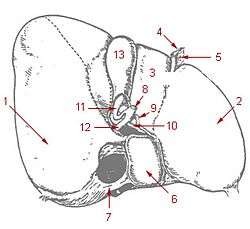 1: Right lobe of liver 2: Left lobe of liver 3: Quadrate lobe of liver 4: Round ligament of liver 5: Falciform ligament 6: Caudate lobe of liver 7: Inferior vena cava 8: Common bile duct 9: Hepatic artery 10: Portal vein 11: Cystic duct 12: Common hepatic duct 13: Gallbladder | |
| Details | |
| Identifiers | |
| Latin | ductus hepaticus communis |
| MeSH | D006500 |
| TA | A05.8.01.061 |
| FMA | 14668 |
| Anatomical terminology | |
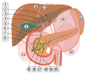
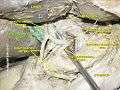
9. Gallbladder, 10–11. Right and left lobes of liver. 12. Spleen.
13. Esophagus. 14. Stomach. 15. Pancreas: 16. Accessory pancreatic duct, 17. Pancreatic duct.
18. Small intestine: 19. Duodenum, 20. Jejunum
21–22. Right and left kidneys.
The front border of the liver has been lifted up (brown arrow).[1]
Clinical significance
The hepatic duct is part of the biliary tract that transports secretions from the liver into the intestines. It carries more volume in people who have had their gallbladder removed.
It is an important anatomic landmark during surgeries such as gall bladder removal. It forms one edge of Calot's triangle, along with the cystic duct and the cystic artery. All constituents of this triangle must be identified to avoid cutting or clipping the wrong structure.
There is some normal anatomic variation of the diameter.
The common hepatic duct is about 6mm in diameter in adults, with some variation.[2] A diameter of more than 8 mm is regarded as abnormal dilatation, and is a sign of cholestasis.[3]
Additional images
 Common hepatic duct
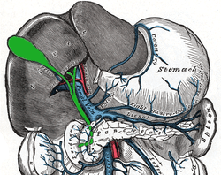 The portal vein and its tributaries.
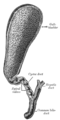 The gall-bladder and bile ducts laid open.
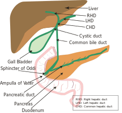
 Common hepatic duct
References
- Standring S, Borley NR, eds. (2008). Gray's anatomy : the anatomical basis of clinical practice. Brown JL, Moore LA (40th ed.). London: Churchill Livingstone. pp. 1163, 1177, 1185–6. ISBN 978-0-8089-2371-8.
- Gray's Anatomy, 39th ed, p. 1228
- Hoeffel, Christine; Azizi, Louisa; Lewin, Maité; Laurent, Valérie; Aubé, Christophe; Arrivé, Lionel; Tubiana, Jean-Michel (2006). "Normal and Pathologic Features of the Postoperative Biliary Tract at 3D MR Cholangiopancreatography and MR Imaging". RadioGraphics. 26 (6): 1603–1620. doi:10.1148/rg.266055730. ISSN 0271-5333.
External links
- Anatomy photo:38:03-0302 at the SUNY Downstate Medical Center - "Stomach, Spleen and Liver: Contents of the Hepatoduodenal Ligament"
- Illustration
| Authority control |
|---|