Kupffer cell
Kupffer cells, also known as stellate macrophages and Kupffer–Browicz cells, are specialized macrophages located in the liver, lining the walls of the sinusoids. They form part of the mononuclear phagocyte system.
| Kupffer cell | |
|---|---|
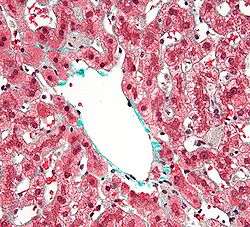 Centrilobular Kupffer cells, with a grey granular cytoplasm, in a resolving liver injury. Liver biopsy. Trichrome stain. | |
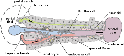 Basic liver structure | |
| Details | |
| Location | Liver |
| Function | Macrophage |
| Identifiers | |
| Latin | macrophagocytus stellatus |
| MeSH | D007728 |
| TH | H3.04.05.0.00016 |
| Anatomical terms of microanatomy | |
Development
Their development begins in the yolk sac where they differentiate into fetal macrophages. Once they enter the blood stream, they migrate to the fetal liver where they stay. There they complete their differentiation into Kupffer cells.[1]
Function
Apart from clearing any bacteria, red blood cells are also broken down by phagocytic action, where the hemoglobin molecule is split. The globin chains are re-used, while the iron-containing portion, heme, is further broken down into iron, which is re-used, and bilirubin, which is conjugated to glucuronic acid within hepatocytes and secreted into the bile.
Helmy et al. identified a receptor present in Kupffer cells, the complement receptor of the immunoglobulin family (CRIg). Mice without CRIg could not clear complement system-coated pathogens. CRIg is conserved in mice and humans and is a critical component of the innate immune system.[2]
Clinical significance
Kupffer cell activation is responsible for early ethanol-induced liver injury, common in chronic alcoholics. Chronic alcoholism and liver injury deal with a two hit system. The second hit is characterized by an activation of the Toll-like receptor 4 (TLR4) and CD14, receptors on the Kupffer cell that internalize endotoxin (lipopolysaccharide or LPS). This activates the transcription of pro-inflammatory cytokines (Tumor necrosis factor-alpha or TNFα) and production of superoxides (a pro-oxidant). TNFα will then enter the stellate cell in the liver, leading to collagen synthesis and fibrosis. Fibrosis will eventually cause cirrhosis, or loss of function of the liver.[3]
History
The cells were first observed by Karl Wilhelm von Kupffer in 1876.[4] The scientist called them "Sternzellen" (star cells or hepatic stellate cell) but thought, inaccurately, that they were an integral part of the endothelium of the liver blood vessels and that they originated from it. In 1898, after several years of research, Tadeusz Browicz identified them, correctly, as macrophages.[5][6]
References
- Naito M, Hasegawa G, Takahashi K (1997). "Development, differentiation, and maturation of Kupffer cells". Microscopy Research and Technique. 39 (4): 350–64. doi:10.1002/(SICI)1097-0029(19971115)39:4<350::AID-JEMT5>3.0.CO;2-L. PMID 9407545.
- Helmy K, Katschke K, Gorgani N, Kljavin N, Elliott J, Diehl L, Scales S, Ghilardi N, van Lookeren Campagne M (2006). "CRIg: a macrophage complement receptor required for phagocytosis of circulating pathogens". Cell. 124 (5): 915–27. doi:10.1016/j.cell.2005.12.039. PMID 16530040.
- Michael D. Wheeler (2004). "Endotoxin and Kupffer Cell Activation in Alcoholic Liver Disease". National Institute on Alcohol Abuse and Alcoholism of the NIH.
- Haubrich W (2004). "Kupffer of Kupffer cells". Gastroenterology. 127 (1): 16. doi:10.1053/j.gastro.2004.05.041. PMID 15236167.
- Szymańska R, Schmidt-Pospuła M (1979). "Studies of liver's reticuloendothelial cells by Tadeusz Browicz and Karl Kupffer. A historical outline". Archiwum historii medycyny. 42 (3): 331–6. PMID 386989.
- Stachura J, Gałazka K (2003). "History and current status of Polish gastroenterological pathology". Journal of Physiology and Pharmacology. 54 Suppl 3: 183–92. PMID 15075472.
External links
- Anatomy photo: digestive/mammal/liver5/liver4 - Comparative Organology at University of California, Davis - "Mammal, liver (EM, Low)"
- Histology image: 15508loa – Histology Learning System at Boston University
- Kupffer Cell Foundation - The mission of the Kupffer cell Foundation is to stimulate and support research and education to improve knowledge on the role of the Kupffer cell and sinusoidal barrier in healthy and diseased liver
| Authority control |
|---|