Hemangiosarcoma
Hemangiosarcoma is a rapidly growing, highly invasive variety of cancer that occurs almost exclusively in dogs, and only rarely in cats, horses, mice,[1] or humans (vinyl chloride toxicity). It is a sarcoma arising from the lining of blood vessels; that is, blood-filled channels and spaces are commonly observed microscopically. A frequent cause of death is the rupturing of this tumor, causing the patient to rapidly bleed to death.
| Hemangiosarcoma | |
|---|---|
| Specialty | Veterinary medicine |
The term "angiosarcoma", when used without a modifier, usually refers to hemangiosarcoma. However, glomangiosarcoma (8710/3) and lymphangiosarcoma (9170/3) are distinct conditions [in humans]. Hemangiosarcomas are commonly associated with toxic exposure to thorium dioxide (Thorotrast), vinyl chloride, and arsenic.
Dogs

Hemangiosarcoma is quite common in dogs, and more so in certain breeds including German Shepherd Dogs and Golden Retrievers.[2] It also occurs in cats, but much more rarely. Dogs with hemangiosarcoma rarely show clinical signs until the tumor has become very large and has metastasized. Typically, clinical signs are due to hypovolemia after the tumor ruptures, causing extensive bleeding. Owners of the affected dogs often discover that the dog has hemangiosarcoma only after the dog collapses.
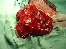
The tumor most often appears on the spleen, right heart base, or liver, although varieties also appear on or under the skin or in other locations. It is the most common tumor of the heart, and occurs in the right atrium or right auricular appendage. Here it can cause right-sided heart failure, arrhythmias, pericardial effusion, and cardiac tamponade. Hemangiosarcoma of the spleen or liver is the most common tumor to cause hemorrhage in the abdomen.[3] Hemorrhage secondary to splenic and hepatic tumors can also cause ventricular arrythmias. Hemangiosarcoma of the skin usually appears as a small red or bluish-black lump. It can also occur under the skin. It is suspected that in the skin, hemangiosarcoma is caused by sun exposure.[3] Occasionally, hemangiosarcoma of the skin can be a metastasis from visceral hemangiosarcoma. Other sites the tumor may occur include bone, kidneys, the bladder, muscle, the mouth, and the central nervous system.
Clinical features
Presenting complaints and clinical signs are usually related to the site of origin of the primary tumor or to the presence of metastases, spontaneous tumor rupture, coagulopathies, or cardiac arrhythmias. More than 50% of patients are presented because of acute collapse after spontaneous rupture of the primary tumor or its metastases. Some episodes of collapse are a result of ventricular arrhythmias, which are relatively common in dogs with splenic or cardiac HSA.[4]
Most common clinical signs of visceral hemangiosarcoma include loss of appetite, arrhythmias, weight loss, weakness, lethargy, collapse, pale mucous membranes, and/or sudden death. An enlarged abdomen is often seen due to hemorrhage. Metastasis is most commonly to the liver, omentum, lungs, or brain.
A retrospective study published in 1999 by Ware, et al., found a 5 times greater risk of cardiac hemangiosarcoma in spayed vs. intact female dogs and a 2.4 times greater risk of hemangiosarcoma in neutered dogs as compared to intact males.. The validity of this study is in dispute. (Personal communication; Modiano, Sackmann)
Clinicopathologic findings
Hemangiosarcoma can cause a wide variety of hematologic and hemostatic abnormalities, including anemia, thrombocytopenia (low platelet count), disseminated intravascular coagulation (DIC); presence of nRBC, schistocytes, and acanthocytes in the blood smear; and leukocytosis with neutrophilia, left shift, and monocytosis.
A definitive diagnosis requires biopsy and histopathology. Cytologic aspirates are usually not recommended, as the accuracy rate for a positive diagnosis of malignant splenic disease is approximately 50%. This is because of frequent blood contamination and poor exfoliation. Surgical biopsy is the typical approach in veterinary medicine.
Symptoms
Dogs rarely show symptoms of hemangiosarcoma until after the tumor ruptures, causing extensive bleeding. Then symptoms can include short-term lethargy, loss of appetite, enlarged abdomen, weakness in the back legs, paled colored tongue and gums, rapid heart rate, and a weak pulse.[5]
Treatments
Treatment includes chemotherapy and, where practical, removal of the tumor with the affected organ, such as with a splenectomy. Splenectomy alone gives an average survival time of 1–3 months. The addition of chemotherapy, primarily comprising the drug doxorubicin, alone or in combination with other drugs, can increase the average survival time to 2–4 months, or more.
A more favorable outcome has been demonstrated in recent research conducted at University of Pennsylvania Veterinary School, in dogs treated with a compound derived from the Coriolus versicolor (commonly known as "Turkey Tail") mushroom:[6]
“We were shocked,” Cimino Brown said. “Prior to this, the longest reported median survival time of dogs with hemangiosarcoma of the spleen that underwent no further treatment was 86 days. We had dogs that lived beyond a year with nothing other than this mushroom as treatment.”There were not statistically significant differences in survival between the three dosage groups, though the longest survival time was highest in the 100 mg group, at 199 days, eclipsing the previously reported survival time.
The results were so surprising, in fact, that the researchers asked Penn Vet pathologists to recheck the dogs’ tissue biopsies to make sure that the dogs really had the disease. “They reread the samples and said, yes, it’s really hemangiosarcoma,” Cimino Brown said. Chemotherapy is available for treating hemangiosarcoma, but many owners opt not to pursue that treatment once their dog is diagnosed.
“It doesn’t hugely increase survival, it’s expensive and it means a lot of back and forth to the vet for the dog,” Cimino Brown said. “So you have to figure in quality of life.”
This treatment does not always work. So, one should always be prepared for their pet to have the same survival time as a dog who is untreated. Visceral hemangiosarcoma is usually fatal even with treatment, and usually within weeks or, at best, months. In the skin, it can be cured in most cases with complete surgical removal as long as there is not visceral involvement.[3]
References
- https://toxnet.nlm.nih.gov/cgi-bin/sis/search/a?dbs+hsdb:@term+@DOCNO+893
- Ettinger, Stephen J.; Feldman, Edward C. (1995). Textbook of Veterinary Internal Medicine (4th ed.). W.B. Saunders Company. ISBN 0-7216-6795-3.
- Morrison, Wallace B. (1998). Cancer in Dogs and Cats (1st ed.). Williams and Wilkins. ISBN 0-683-06105-4.
- Nelson; et al. (2005). Manual of Small Animal Internal Medicine. St. Louis, Missouri: Elsevier Mosby.
- Farricelli, Adrienne. "The Silent Killer: Hemangiosarcomas or a Ruptured, Bleeding Spleen in Dogs". PetHelpful. Retrieved 23 March 2019.
- "Compound Derived From a Mushroom Lengthens Survival Time in Dogs With Cancer, Penn Vet Study Finds". University of Pennsylvania.
External links
| Classification |
|---|
- "Flat-Coated Retriever Hemangiosarcoma". AKC.org.
- "Heart hemangiosarcoma in Cats and Dogs". Pet Cancer Center.
- "Hemangiosarcoma". Canine Cancer Awareness. - includes links to current studies
- "Hemangiosarcoma". Cure Hemangiosarcoma. - includes links to current and past studies of hemangiosarcoma
- "Hemangiosarcoma". Mar Vista Animal Medical Center.
- "Hemangiosarcoma". Vetinfo.
- "Hemangiosarcoma in Cats and Dogs". Pet Cancer Center.
- "Hemangiosarcoma research". Modiano Lab of the Univ. of Minnesota. - includes links to other canine cancer research
- "Vet Corner: Hemangiosarcoma". Adopt a Golden Retriever.