Bowman's capsule
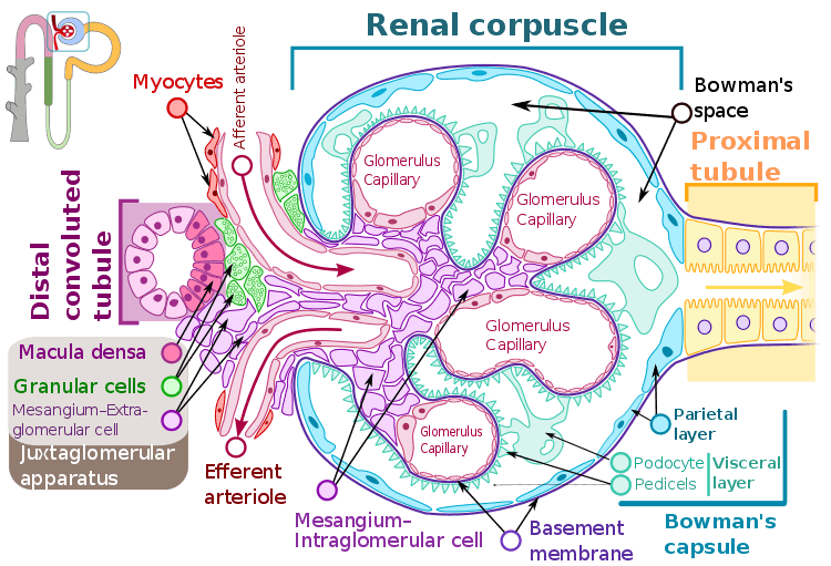
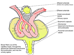
Bowman's capsule (or the Bowman capsule, capsula glomeruli, or glomerular capsule) is a cup-like sack at the beginning of the tubular component of a nephron in the mammalian kidney that performs the first step in the filtration of blood to form urine. A glomerulus is enclosed in the sac. Fluids from blood in the glomerulus are collected in the Bowman's capsule .
| Bowman's capsule | |
|---|---|
 Glomerulus. (Bowman's capsule not labeled, but visible at top.) | |
| Details | |
| Precursor | Metanephric blastema |
| Location | Nephron of kidney |
| Identifiers | |
| Latin | capsula glomeruli |
| MeSH | D050476 |
| FMA | 15626 |
| Anatomical terminology | |
Structure
Outside the capsule, there are two "poles":
- The vascular pole is the side with the afferent arteriole and efferent arteriole.
- The urinary pole is the side with the proximal convoluted tubule.
Inside the capsule, the layers are as follows, from outside to inside:
- Parietal layer—A single layer of simple squamous epithelium. Does not function in filtration.
- Bowman's space (or "urinary space", or "capsular space")—Between the visceral and parietal layers, into which the filtrate enters after passing through the filtration slits.[1]
- Visceral layer—Lies just above the thickened glomerular basement membrane and is made of podocytes. Beneath the visceral layer lie the glomerular capillaries.
- Filtration barrier—The filtration barrier is composed of the fenestrated endothelium of the glomerular capillaries, the fused basal lamina of the endothelial cells and podocytes, and the filtration slits of the podocytes. The barrier permits the passage of water, ions, and small molecules from the bloodstream into the Bowman's space. The barrier prevents the passage of large and/or negatively charged proteins (such as albumin). The basal lamina of the filtration barrier is composed of three layers. The first layer is the lamina rara externa, adjacent to the podocyte processes. The second layer is the lamina rara interna, adjacent to the endothelial cells. The final layer is the lamina densa which is a darker central zone of the basal lamina. It consists of the meshwork of type IV collagen and laminin which act as a selective macromolecular filter.
Function
The process of filtration of the blood in the Bowman's capsule is ultrafiltration (or glomerular filtration), and the normal rate of filtration is 125 ml/min, equivalent to 80 times the daily blood volume.
Any proteins under roughly 30 kilodaltons can pass freely through the membrane, although there is some extra hindrance for negatively charged molecules due to the negative charge of the basement membrane and the podocytes.
Any small molecules such as water, glucose, salt (NaCl), amino acids, and urea pass freely into Bowman's space, but cells, platelets and large proteins do not.
As a result, the filtrate leaving the Bowman's capsule is very similar to blood plasma (filtrate or glomerular filtrate is composed of blood plasma minus plasma protein i.e. it contains all the components of blood plasma except the proteins) in composition as it passes into the proximal convoluted tubule.
Clinical significance
Measuring the glomerular filtration rate (GFR) is a diagnostic test of kidney function.[2]
A decreased GFR may be a sign of renal failure.
A number of diseases can result in various problems within the glomerulus. Examples include acute proliferative (endocapillary) glomerulonephritis, mesangioproliferative glomerulonephritis, mesangiocapillary (membranoproliferative) glomerulonephritis, acute crescentic glomerulonephritis, focal segmental glomerulonephritis, and diabetic glomerulosclerosis.
History
Bowman's capsule is named after Sir William Bowman (1816–1892), a British surgeon and anatomist.[3] However, thorough microscopical anatomy of kidney including the nephronic capsule was first described by Ukrainian surgeon and anatomist from the Russian Empire, Prof. Alexander Schumlansky (1748–1795), in his 1782 doctoral thesis "De structura renum" ("About Kidney Structure", in Latin); thus, much prior to Bowman.[4]
Together with the glomerulus it is known as a renal corpuscle, or a Malpighian corpuscle, named after Marcello Malpighi (1628–1694), an Italian physician and biologist. This name is not used widely anymore, probably to avoid confusion with Malpighian bodies of the spleen.
See also
Additional images
 Glomerulus.
References
- Histology image:22401lba from Vaughan, Deborah (2002). A Learning System in Histology: CD-ROM and Guide. Oxford University Press. ISBN 978-0195151732.
- Romagnani, Paola; Anders, Hans-Joachim (2019). "Excretory System". In Brüne, Martin; Schiefenhövel, Wulf (eds.). Oxford Handbook of Evolutionary Medicine. Oxford University Press. ISBN 978-0198789666.
- Bowman, William; Royal Society of London. Philosophical transactions, v. 32, p. 57-80, 1842 (1842). On the structure and use of the Malpighian bodies of the kidney: with observations on the circulation through that gland. London: Taylor.
- Schumlansky, Aleksander (1782). Dissertatio Inauguralis Anatomica De Structura Renum Quam Pro Licentia Summos In Medicina Honores Et Privilegia Doctoralia Legitime Obtinendi In Inclyta Argentoratensium Universitate Solenni Eruditorum Examini Submittit Alexander Schumlansky Poltawo-Russus Die XVI. Novembr. A. MDCCLXXXII (in Latin). Argentorati [Strasbourg]. p. 92.
External links
- Histology image: 16006loa – Histology Learning System at Boston University
- Diagram at ircc.edu