Enamel hypoplasia
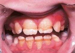
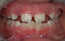
Enamel hypoplasia is a defect of the teeth in which the enamel is deficient in amount,[1] caused by defective enamel matrix formation. Defects are commonly split into one of four categories, pit-form, plane-form, linear-form, and localised enamel hypoplasia.[2][3][4] In many cases the enamel crown has pits or a groove on it, and in extreme cases, sections of the tooth have no enamel, exposing the dentin.[5] Enamel hypoplasia varies substantially among populations and can be used to infer health and behaviour in past populations. Defects have also been found in a variety of non-human animals.[6][7][8]
| Enamel hypoplasia | |
|---|---|
 | |
| Specialty | Dentistry |
Causes
It can be caused by any of the following:
- Nutritional factors.[12]
- Some diseases (such as undiagnosed and untreated celiac disease,[13][14][15] chicken pox, congenital syphilis[12]).
- Hypocalcemia.[12]
- Fluoride ingestion (dental fluorosis).[12]
- Birth injury.[12]
- Preterm birth.[12]
- Infection.[12]
- Trauma from a deciduous tooth.[12]
- Abnormalities in amelogenesis: Enamel hypoplasia (EH) is a developmental defect that can affect the primary and permanent teeth in one of two ways. It is sometimes identified as a physically missing tooth structure, and can be seen as pits, grooves or just missing parts in the crown of the tooth. Hypomineralization, on the other hand, is a mere decrease in the mineral content of the enamel. It can be severe enough to give the tooth a translucent appearance or mild enough to maintain its opacity. It is hypomineralization that leads to soft enamel. Enamel hypocalcification is a defect of tooth enamel in which normal amounts of enamel are produced but are hypomineralized. In this defect the enamel is softer than normal.[16] Enamel hypoplasia is a defect involving the surface of the enamel associated with a reduced localized thickness of enamel without dentinal exposure. Studies have suggested that enamel hypoplasia, particularly in anterior teeth, is associated with early childhood caries. Hypoplasia in primary anterior teeth is associated with prenatal smoking, excessive weight gain, low birth weight, prematurity and post-natal measles.[17] The random distribution of the round shape of the hypoplastic defects of enamel with hypomineralization, in which there is a continuity of the incremental lines, suggested the possibility that some ameloblasts maintained hypoactivity of their secretory function during their total life span.[18] These changes were confined to the ameloblastic layer, but in severe cases, there was an associated change in the stratum intermedium. They may be submitted as evidence that enamel hypoplasia is essentially a disturbance in the ameloblasts.[19] Enamel hypoplasia or hypo mineralization may be caused by hereditary factors and environmental factors including
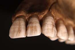 Teeth displaying enamel hypoplasia lines, linear defects of enamel that form during crowns development as a result of periods of nutritional stress or disease during infancy and childhood.
- systemic factors such as nutritional factors, exanthematous diseases like measles and chickenpox, congenital syphilis, hypocalcemia, birth injury or premature birth, fluoride ingestion or idiopathic causes.
- local factors such as infection or trauma from a deciduous tooth. Hereditary enamel hypoplasia/hypomineralization is known as amelogenesis imperfecta. It is transmitted in the family as a mendelian dominant trait which affects enamel of all the teeth, deciduous as well as permanent.[20] Environmental enamel hypoplasia/hypomineralization of systemic or local origin is also termed as “chronologic hypoplasia”. This lesion is found in areas of those teeth where the enamel was being formed during the systemic or local disturbance. Since the formation of enamel extends over a long period and the systemic or local disturbance, in most cases are of short duration, the defect is limited to a circumscribed area of the affected teeth or tooth. Thus knowing the chronological development of deciduous and permanent teeth will make it possible to determine from the location of the defect, the approximate time at which the injury occurred.[21] More recently enamel hypoplasia has been produced in sheep by
- physical trauma[22][23]
- systemic illness induced by intestinal parasites[24][25]
- a daily high dose of fluoride for a short period[26] These findings indicate that hypoplastic defects are formed during the secretory phase of amelogenesis. ie duration of the insult is relatively short, and it is the severity that determines the extent of the defect and the translucency of the partially formed enamel. Aetiology does not seem to be of major importance, since local and systemic factors result in defects with a similar appearance and physical characteristics.
Turner's hypoplasia
Turner's hypoplasia is an abnormality found in teeth. Its appearance is variable, though usually is manifested as a portion of missing or diminished enamel on permanent teeth. Unlike other abnormalities which affect a vast number of teeth, Turner's hypoplasia usually affects only one tooth in the mouth and, it is referred to as a Turner's tooth.
If Turner's hypoplasia is found on a canine or a premolar, the most likely cause is an infection that was present when the primary (baby) tooth was still in the mouth. Most likely, the primary tooth was heavily decayed and an area of inflamed tissues around the root of the tooth (called a periapical inflammation), affecting the development of the permanent tooth. The appearance of the abnormality will depend on the severity and longevity of the infection.
If Turner's hypoplasia is found in the front (anterior) area of the mouth, the most likely cause is a traumatic injury to a primary tooth. The traumatized tooth, which is usually a maxillary central incisor, is pushed into the developing tooth underneath it and consequently affects the formation of enamel. Because of the location of the permanent tooth's developing tooth bud in relation to the primary tooth, the most likely affected area on the permanent tooth is the facial surface (the side closer to the lips or cheek). White or yellow discoloration may accompany Turner's hypoplasia. Enamel hypoplasia may also be present.
Turner's hypoplasia usually affects the tooth enamel if the trauma occurs prior to the third year of life. Injuries occurring after this time are less likely to cause enamel defects since the enamel is already calcified.
The same type of injury is also associated with the dilaceration of a tooth.
References
- Ash, Major M., Jr. & Nelson, S.J (2003). Dental anatomy, physiology, and occlusion (8th ed.). Philadelphia: W.B. Saunders. ISBN 978-0-7216-9382-8.CS1 maint: multiple names: authors list (link)
- Towle, Ian; Irish, Joel D. (2019). "A probable genetic origin for pitting enamel hypoplasia on the molars of Paranthropus robustus". Journal of Human Evolution. 129: 54–61. doi:10.1016/j.jhevol.2019.01.002. PMID 30904040.
- Hillson, Simon; Bond, Sandra (1997). "Relationship of enamel hypoplasia to the pattern of tooth crown growth: A discussion". American Journal of Physical Anthropology. 104 (1): 89–103. doi:10.1002/(SICI)1096-8644(199709)104:1<89::AID-AJPA6>3.0.CO;2-8. PMID 9331455.
- Skinner, Mark F.; Skinner, Matthew M.; Pilbrow, Varsha C.; Hannibal, Darcy L. (2016). "An Enigmatic Hypoplastic Defect of the Maxillary Lateral Incisor in Recent and Fossil Orangutans from Sumatra (Pongo abelii) and Borneo (Pongo pygmaeus)" (PDF). International Journal of Primatology. 37 (4–5): 548–567. doi:10.1007/s10764-016-9920-2.
- Groote, Isabelle De; Irish, Joel D.; Dove, Eleanor R.; Towle, Ian (2018). "Severe Plane-Form Enamel Hypoplasia in a Dentition from Roman Britain" (PDF). Dental Anthropology Journal. 30: 16–24. doi:10.26575/daj.v30i1.23.
- Dobney, Keith; Ervynck, Anton (2000). "Interpreting Developmental Stress in Archaeological Pigs: The Chronology of Linear Enamel Hypoplasia". Journal of Archaeological Science. 27 (7): 597–607. doi:10.1006/jasc.1999.0477.
- Towle, Ian; Irish, Joel D.; De Groote, Isabelle (2018). "Amelogenesis imperfecta in the dentition of a wild chimpanzee" (PDF). Journal of Medical Primatology. 47 (2): 117–119. doi:10.1111/jmp.12323. PMID 29112236.
- Moggi-Cecchi, Jacopo; Crovella, Sergio (1991). "Occurrence of Enamel Hypoplasia in the Dentitions of Simian Primates". Folia Primatologica. 57 (2): 106–110. doi:10.1159/000156571. PMID 1786905.
- "Diagnosis of Celiac Disease". National Institute of Health (NIH). Archived from the original on 15 May 2017. Retrieved 6 June 2017.CS1 maint: BOT: original-url status unknown (link)
- Dental Enamel Defects and Celiac Disease Archived March 5, 2016, at the Wayback Machine National Institute of Health (NIH)
- Pastore L, Carroccio A, Compilato D, Panzarella V, Serpico R, Lo Muzio L (2008). "Oral manifestations of celiac disease". J Clin Gastroenterol (Review). 42 (3): 224–32. doi:10.1097/MCG.0b013e318074dd98. hdl:10447/1671. PMID 18223505.
- Krishan, Kewal; Garg, Arunk; Kanchan, Tanuj; Machado, Meghna; Rao, Ashwin (2015). "Enamel hypoplasia and its role in identification of individuals: A review of literature". Indian Journal of Dentistry. 6 (2): 99–102. doi:10.4103/0975-962X.155887. PMC 4455163. PMID 26097340.
- Dental Enamel Defects and Celiac Disease Archived 2016-03-05 at the Wayback Machine National Institute of Health (NIH)
- Ferraz, E. G.; Campos Ede, J.; Sarmento, V. A.; Silva, L. R. (2012). "The oral manifestations of celiac disease: Information for the pediatric dentist". Pediatric Dentistry. 34 (7): 485–8. PMID 23265166.
- Giuca, M. R.; Cei, G.; Gigli, F.; Gandini, P. (2010). "Oral signs in the diagnosis of celiac disease: Review of the literature". Minerva Stomatologica. 59 (1–2): 33–43. PMID 20212408.
- "Enamel Hypoplasia and Hypomineralization | Colgate® Oral Care".
- https://www.ada.org/~/media/ADA/Education%20and%20Careers/Files/6_Fontana_QUEST%20Enamel%20Hypoplasia%20Project.pdf
- Koshiba, H.; Kimura, O.; Nakata, M. (1977). "A clinical and histologic observation of enamel hypoplasia in a case of epidermolysis bullosa hereditaria". Oral Surgery, Oral Medicine, and Oral Pathology. 43 (4): 585–9. doi:10.1016/0030-4220(77)90113-X. PMID 265487.
- Kerr, Donald A. (1944). "Histological changes in the enamel organ responsible for enamel hypoplasia". American Journal of Orthodontics and Oral Surgery. 30 (11): 673–679. doi:10.1016/S0096-6347(44)90066-9.
- Rajendran, R (2009). "Developmental Disturbances of Oral and para oral structures". In Rajendran, R; Sivapathasundaram, B (eds.). Shafer's Textbook of Oral Pathology. pp. 3–79.
- Kumar, G. (2011). Orban's Oral Histology and Embryology (13th ed.). India: Elsevier. pp. 72–87.
- Suckling, G. (1980). "Defects of Enamel in Sheep Resulting from Trauma During Tooth Development". Journal of Dental Research. 59 (9): 1541–1548. doi:10.1177/00220345800590092701. PMID 6931141.
- Suckling, G.W.; Purdell-Lewis, D.J. (1982). "The Pattern of Mineralization of Traumatically-induced Developmental Defects of Sheep Enamel Assessed by Microhardness and Microradiography". Journal of Dental Research. 61 (10): 1211–1216. doi:10.1177/00220345820610102201. PMID 6956605.
- Suckling, Grace; Elliott, D.C.; Thurley, D.C. (1983). "The production of developmental defects of enamel in the incisor teeth of penned sheep resulting from induced parasitism". Archives of Oral Biology. 28 (5): 393–399. doi:10.1016/0003-9969(83)90134-6. PMID 6578757.
- Suckling, Grace; Elliott, D.C.; Thurley, D.C. (1986). "The macroscopic appearance and associated histological changes in the enamel organ of hypoplastic lesions of sheep incisor teeth resulting from induced parasitism". Archives of Oral Biology. 31 (7): 427–439. doi:10.1016/0003-9969(86)90016-6. PMID 3467666.
- Suckling, Grace; Thurley, D.C. (1984). "Histological, macroscopic and microhardness observations of fluoride-induced changes in the enamel organ and enamel of sheep incisor teeth". Archives of Oral Biology. 29 (3): 165–177. doi:10.1016/0003-9969(84)90050-5. PMID 6587836.