

Tuberculosis Photos
 ShareCompartir
One of the Non-routine Vaccines by Disease
WARNING: Some of these photos might be unsuitable for children. Viewing discretion is advised.
Photos of the Disease
From the Public Health Image Library
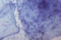
Photo ID#5790
This photomicrograph reveals Mycobacterium tuberculosis bacteria using acid-fast Ziehl-Neelsen stain; Magnified 1000X.
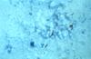
Photo ID# 4427
This photomicrograph reveals Mycobacterium tuberculosis bacteria using acid-fast Ziehl-Neelsen stain; Magnified 1000X.
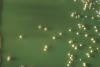
Photo ID# 2904
Here a Lowenstein-Jensen plate culture has been inoculated with 15 strains of Mycobacteria spp..

Photo ID# 2127
A photomicrograph of Mycobacterium tuberculosis bacteria from a sputum specimen, and viewed with Ziehl-Neelsen stain.

Photo ID#2128
Photomicrograph of a sputum sample containing Mycobacterium tuberculosis.
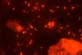
Photo ID# 6468
M. tuberculosis in a sputum smear is stained using fluorescent auramine with acridine orange counterstain; Mag.-950x.
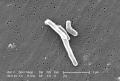
Photo ID# 8436
At a magnification of 13172x, this scanning electron micrograph (SEM) depicted a number of Gram-positive Mycobacterium tuberculosis bacteria.
Images of People Affected by the Disease
From the Public Health Image Library
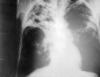
Photo ID# 2543
An anteroposterior X-ray of a patient diagnosed with advanced bilateral pulmonary tuberculosis.
- Page last reviewed: May 23, 2011
- Page last updated: May 23, 2011
- Content source: