


Transmission
 ShareCompartir
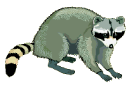
The Infectious Path of the Rabies Virus
- Raccoon is bitten by a rabid animal.
- Rabies virus enters the raccoon through infected saliva.
- Rabies virus spreads through the nerves to the spinal cord and brain.
- The virus incubates in raccoon’s body for apporximately 3-12 weeks. The raccoon has no signs of illness during this time.
- When it reaches the brain, the virus multiplies rapidly, passes to the salivary glands, and the raccoon begins to show signs of the disease.
- The infected animal usually dies within 7 days of becoming sick.
Following primary infection, the virus enters an eclipse phase in which it cannot be easily detected within the host. This phase may last for several days or months. Investigations have shown both direct entry of virus into peripheral nerves at the site of infection and indirect entry after viral replication in nonnervous tissue (i.e., muscle cells).
During the eclipse phase, the host immune defenses may confer cell-mediated immunity against viral infection because rabies virus is a good antigen. The uptake of virus into peripheral nerves is important for progressive infection to occur (number 3 above).
After uptake into peripheral nerves, rabies virus is transported to the central nervous system (CNS) via retrograde axoplasmic flow. Typically this occurs via sensory and motor nerves at the initial site of infection. The incubation period (number 4 above) is the time from exposure to onset of clinical signs of disease. The incubation period may vary from a few days to several years, but is typically 1 to 3 months. Dissemination of virus within the CNS is rapid, and includes early involvement of limbic system neurons (number 5 above).
Active cerebral infection is followed by passive centrifugal spread of virus to peripheral nerves. The amplification of infection within the CNS occurs through cycles of viral replication and cell-to-cell transfer of progeny virus. Centrifugal spread of virus may lead to the invasion of highly innervated sites of various tissues, including the salivary glands. During this period of cerebral infection, the classic behavioral changes associated with rabies develop.
Pathology
Pathology of rabies infection is typically defined by encephalitis and myelitis. Perivascular infiltration with lymphocytes, polymorphonuclear leukocytes, and plasma cells can occur throughout the entire CNS. Rabies infection frequently causes cytoplasmic eosinophilic inclusion bodies (Negri bodies) in neuronal cells, especially pyramidal cells of the hippocampus and Purkinje cells of the cerebellum. These inclusions have been identified as areas of active viral replication by the identification of rabies viral antigen.
Several factors may affect the outcome of rabies exposure. These include the virus variant, the dose of virus inoculum, the route and location of exposure, as well as individual host factors, such as age and host immune defenses.
Related Links
- Page last reviewed: June 30, 2011
- Page last updated: June 30, 2011
- Content source: