


Case #446 - June, 2017
 ShareCompartir
Blood specimens were collected at night from members of a village in Myanmar endemic for lymphatic filariasis. Roughly one-third of the cases were symptomatic, with or without recurrent episodes of fever and various degrees of lymphedema. The objects in Figures A and B were observed on Giemsa-stained thick blood films from a few of the asymptomatic cases at 500x oil magnification; Figures C and D show close detail of the object in Figure B at 1000x oil magnification. The objects measured on average 200 micrometers in length. What is your diagnosis? Based on what criteria?
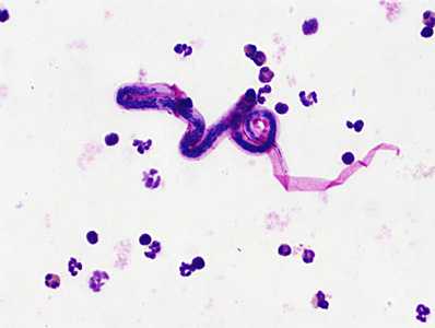
Figure A
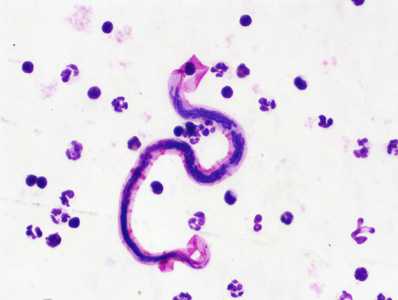
Figure B
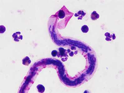
Figure C
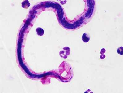
Figure D
Case Answer
This was a case of lymphatic filariasis caused by Brugia malayi. Diagnostic morphologic features included:
- microfilaria whose sheaths stained pink with Giemsa and within the size range for B. malayi (175-230 micrometers)
- dense nuclear column in which individual nuclei are easily defined
- microfilariae with a relatively long head space and a tail with terminal and subterminal nuclei (Figure D)
More on lymphatic filariasis
Images presented in the monthly case studies are from specimens submitted for diagnosis or archiving. On rare occasions, clinical histories given may be partly fictitious.
DPDx is an education resource designed for health professionals and laboratory scientists. For an overview including prevention and control visit www.cdc.gov/parasites/.
- Page last reviewed: July 19, 2017
- Page last updated: July 19, 2017
- Content source:
- Global Health – Division of Parasitic Diseases and Malaria
- Notice: Linking to a non-federal site does not constitute an endorsement by HHS, CDC or any of its employees of the sponsors or the information and products presented on the site.
- Maintained By: