


Case #444 - May, 2017
 ShareCompartir
A 37-year-old male immigrant from the Middle East sought medical attention for pain in the upper right abdominal quadrant. Ultrasonography revealed a single compartment hepatic cyst approximately 7 cm in diameter and during the imaging, a fine biopsy needle was used to obtain an aspirate specimen. The specimen was sent to the CDC DPDx Team for examination. Figures A–D show what was observed at 200x magnification; Figures A and B are of the same microscopic field but different focal planes. What is your diagnosis? Based on what criteria? What, if any, other tests would you recommend?
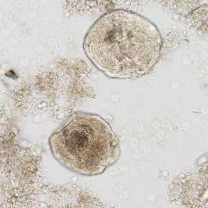
Figure A
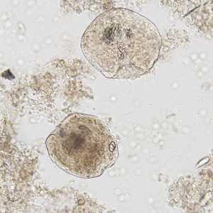
Figure B
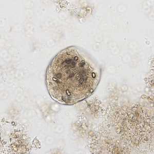
Figure C
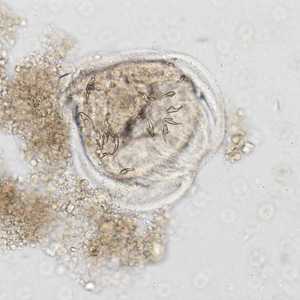
Figure D
Case Answer
This was a case of hydatid disease (human echinococcosis) caused by the larval stage of Echinococcus granulosus. Morphologic features shown in the Figures included:
- Multiple protoscoleces, identifiable by the refractile hooks, observed in the aspirated fluid.
Careful analysis of imaging techniques coupled with serologic testing is the preferred course of diagnosing infection with Echinococcus to minimize the chance of the cyst rupturing and spreading the infection to surrounding areas and/or organs.
Dogs and other canids are the definitive host for Echinococcus spp.; humans are only infected with the larvae stage (protoscoleces) after ingestion of food or water contaminated with eggs from infected dogs (or other canids).
More on Echinococcosis
Images presented in the monthly case studies are from specimens submitted for diagnosis or archiving. On rare occasions, clinical histories given may be partly fictitious.
DPDx is an education resource designed for health professionals and laboratory scientists. For an overview including prevention and control visit www.cdc.gov/parasites/.
- Page last reviewed: July 19, 2017
- Page last updated: July 19, 2017
- Content source:
- Global Health – Division of Parasitic Diseases and Malaria
- Notice: Linking to a non-federal site does not constitute an endorsement by HHS, CDC or any of its employees of the sponsors or the information and products presented on the site.
- Maintained By: