


Case #442 - April, 2017
 ShareCompartir
A 6-year-old child from a community in rural southeastern United States presented with severe upper right abdominal pain, fever, chills, decreased appetite and weight loss. The child expired 2 weeks after being admitted for symptoms that included hepatitis. At autopsy, a liver biopsy was obtained and processed. Figures A and B show what was observed on a hematoxylin and eosin (H & E) stained liver tissue section at 100x and 500x oil magnification respectively. What is your diagnosis? Based on what criteria?
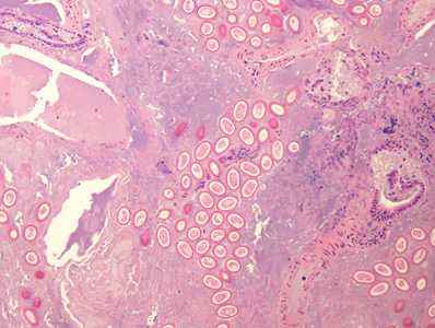
Figure A
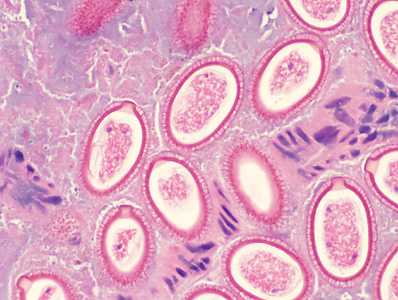
Figure B
Case Answer
This was a case of hepatic capillariasis, a rare but serious illness that is most often found at autopsy. Morphologic features shown in the figures were:
- Numerous eggs located in the parenchyma of the liver.
- Oval-shaped eggs with a thick striated shell and polar prominences consistent with C. hepatica.
More on: hepatic capillariasis
Images presented in the monthly case studies are from specimens submitted for diagnosis or archiving. On rare occasions, clinical histories given may be partly fictitious.
DPDx is an education resource designed for health professionals and laboratory scientists. For an overview including prevention and control visit www.cdc.gov/parasites/.
- Page last reviewed: May 11, 2017
- Page last updated: May 17, 2017
- Content source:
- Global Health – Division of Parasitic Diseases and Malaria
- Notice: Linking to a non-federal site does not constitute an endorsement by HHS, CDC or any of its employees of the sponsors or the information and products presented on the site.
- Maintained By: