


Case #421 - June 2016
 ShareCompartir
A 62-year-old female from rural Manitoba Province, Canada presented to a health care provider with abdominal pain, diarrhea, and peripheral eosinophilia. A stool specimen was collected in sodium acetate-acetic acid-formalin (SAF) and a formalin-ethyl acetate (FEA) concentration was performed. Figures A-D show what was observed microscopically on a wet mount; Figures C and D were stained with iodine. What is your diagnosis? Based on what criteria?
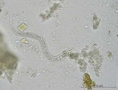
Figure A
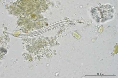
Figure B
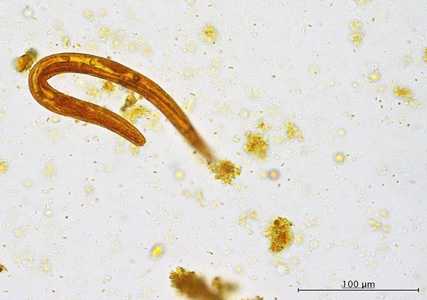
Figure C
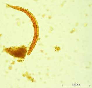
Figure D
Case Answer
This was a case of strongyloidiasis caused by Strongyloides stercoralis. Diagnostic morphologic features shown in the images included:
- a prominent genital primordium (black arrow, Figures B and C).
- a short buccal canal (red arrow, Figure C).
- a rhabditoid esophagus (blue arrow, Figure B).
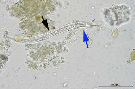
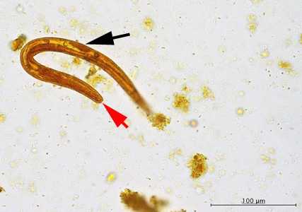
The morphologic features of the larvae shown in the images were consistent with first-stage rhabditiform (L1) larvae which is the stage usually found in stool.
More on: Strongyloidiasis
Acknowledgements
This case and images were kindly provided by Cadham Provinical Public Health Laboratory, University of Manitoba, Winnipeg, Canada.
DPDx is an education resource designed for health professionals and laboratory scientists. For an overview including prevention and control visit www.cdc.gov/parasites/.
- Page last reviewed: August 24, 2016
- Page last updated: August 24, 2016
- Content source:
- Global Health – Division of Parasitic Diseases and Malaria
- Notice: Linking to a non-federal site does not constitute an endorsement by HHS, CDC or any of its employees of the sponsors or the information and products presented on the site.
- Maintained By: