


Case #411 - January 2016
 ShareCompartir
Case Description
A 23-year-old female with no documented travel history presented with iron deficiency anemia and periodic abdominal pain. Ova-and-parasite (O&P) examinations of stool were negative. A colonoscopy was performed and a worm-like object (Figures A and B) was observed attached to the mucosa of the ascending colon. The object was removed, sent to Pathology, and processed by routine histologic work-up. Figures C-E show what was observed by the pathologist after sectioning and staining with hematoxylin-and-eosin (H&E). Images were captures and sent to the CDC-DPDx for diagnostic assistance. What is your diagnosis? Based on what criteria?
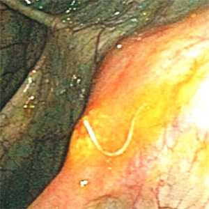
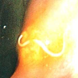
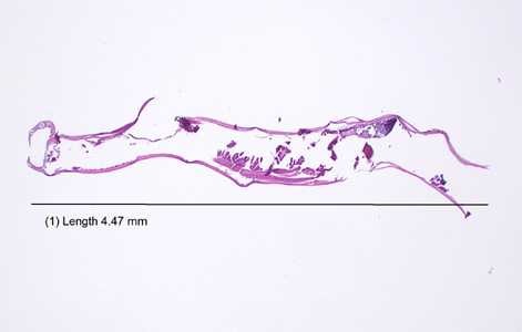
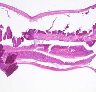
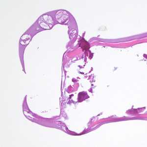
Figure A
Figure B
Figure C
Figure D
Figure E
Case Answer
This was a case of hookworm infection, caused by either Necator americanus or Ancylostoma duodenale. Diagnostic morphologic features included:
- platymyarian somatic musculature (MU, Figure D)
- multi-nucleated intestinal cells with a brush border (IN, Figure D)
- the presence of a copulatory bursa (Figure E), indicating the specimen was a male. The bursa, as presented in these images, is suggestive of Ancylostoma, being wider than long.
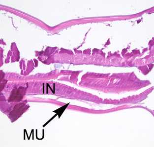
Figure D
More on: hookworm
Answer Figure D
Acknowledgements
This case and images were kindly provided by St. David’s South Austin Medical Center, Austin, TX.
DPDx is an education resource designed for health professionals and laboratory scientists. For an overview including prevention and control visit www.cdc.gov/parasites/.
- Page last reviewed: August 24, 2016
- Page last updated: August 24, 2016
- Content source:
- Global Health – Division of Parasitic Diseases and Malaria
- Notice: Linking to a non-federal site does not constitute an endorsement by HHS, CDC or any of its employees of the sponsors or the information and products presented on the site.
- Maintained By: